Biomedical Engineering Reference
In-Depth Information
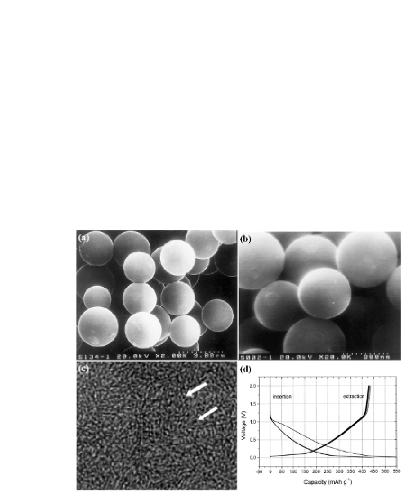
from 100 nm to 5 μm can be obtained by this method [24-26].
In the transmission electron microscopy (TEM) image shown in
Fig. 6.2, a large quantity of uniform nanopores (ca. 0.4 nm) can be
observed distributing in monodispersed hard carbon spherules,
which make the material has a BET specific surface area of
400 m
,
which might be partially attributed to the tiny nanopores inside of
HCSs. It has been reported that similar HCSs can also be prepared
from other materials, such as glucose, starch and rice
carbohydrates [27, 28]. These findings substantially widen
the system category of carbon-based anode materials toward
mass storage.
2
g
-
1
, and can reversibly store lithium up to 430 mAh g
-
1
Figure
Results of electronic microscopy characterization and
electrochemical test of as-prepared HCSs. (a) Scanning electron microscopy
image of monodispersed HCSs with particle size of 5 μm. (b) Scanning
electron microscopy image of monodispersed HCSs with particle size of 1
μm. (c) High-resolution transmission electron microscopy (HRTEM) image
of the interior structure of an HCS, in which micropores can be clearly seen.
(d) Typical plots of voltage vs. capacity of the HCSs as negative electrode in
an Li/HCS cell during the first 10 charge/discharge cycles (all reproduced
with permission from [24],
copyright 2001, Elsevier Science Ltd.).
6.2


Search WWH ::

Custom Search