Biology Reference
In-Depth Information
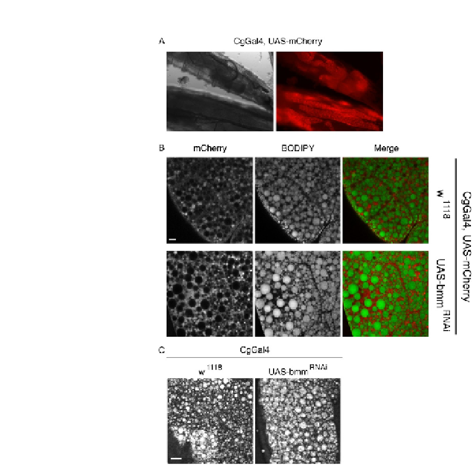
FIGURE 4.2
Imaging LDs in Drosophila larval fat bodies. (A) Live third instar larvae of the CgGal4,
UAS-mCherry strain was imaged directly under a standard fluorescent stereoscope. Left
image shows the bright-field image of larvae and right image shows fat bodies labeled with
mCherry. (B) Flies with bmm knockdown (bottom panels) in the larval fat body have
larger LDs than control (w
1118
, top panels). LDs were stained with BODIPY493/503 in fixed
larval fat bodies and imaged under the LSM 710 confocal microscope. Fat bodies were
labeled with mCherry. (C) LD morphology was imaged under an Olympus two-photon
microscope using the coherent anti-Stokes Raman scattering function. Fresh fat bodies were
imaged in a label-free manner for LDs. Scale bars
¼
10
m
m in (B) and 20
m
m in (C).
(See color plate.)