Biology Reference
In-Depth Information
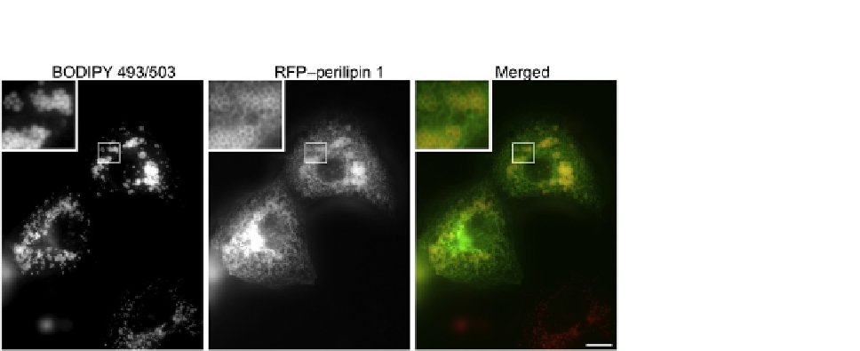
FIGURE 12.3
BODIPY 493/503 stains fat droplets in living cells. COS7 cells were transfected to express a
red fluorescent protein/perilipin 1 fusion as described previously (
Skinner, Harris, Shew,
Abumrad, & Wolins, 2013
) 18 h later, 2
g/ml of BODIPY 493/503 and 1.8 mM fatty acid
were added to the media. After a 4-h incubation, the coverslip was rinsed in phosphate-
buffered saline and imaged. For clearer presentation, in the merged image the green
fluorescing BODIPY 493/503 is shown as red, and the red fluorescent protein/perilipin 1
fusion is shown as green.
m
Add DAPI along with the primary antibodies, the secondary fluorochrome-antibody
conjugates or in the mounting media at 0.5
g/ml.
m
12.2.7.1
Fat droplets in dispersed muscle fibers
We have adapted this protocol for collagenase dispersed skeletal and cardiomyocytes.
We fix and stain these myocytes in suspension. Excess dye and antibodies are washed
away by low speed centrifugation of cells (200
g
for 5 min), removal of supernatant
and resuspension of myocytes in PBS, five times after fixation, and five times after
addition of primary antibodies and after fluorochrome-conjugated antibodies. After
the final wash we resuspend the myocytes in elvanol, pipette the elvanol-myocyte
suspension onto a slide, and cover the suspension with a coverslip. If appropriate,
vital staining of live cells or muscle fibers can be imaged without fixation (
Figs. 12.3
and 12.4
).
12.3
DISCUSSION
12.3.1
Validation of signals
The microscopy techniques for imaging fat droplets and associated cellular structures
described herein employ antibody-based labeling of fat droplet protein coats and the
direct fluorophore staining of the fat droplet core. Immunofluorescence-based