Biology Reference
In-Depth Information
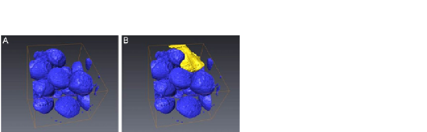
FIGURE 8.6
3D segmentation of a subvolume. (A) Segmentation of all LDs in the region of interest was
performed and represented (blue) and (B), one single mitochondrian was selected to be
segmented (yellow).
It is estimated that 33.2% of this subvolume is filled by LDs. One mitochondrian is
segmented in
Fig. 8.6
B to illustrate the spatial arrangement and interactions between
these two organelles and the change of mitochondria morphology due to the presence
of large volume of LDs in this particular heart muscle specimen.
Although we are still in the process of improving the sample preparation method
for FIB-SEM 3D analysis, we demonstrate the feasibility of studying LD fusions and
LD-mitochondria interactions in cardiac muscle using FIB-SEM 3D imaging tech-
niques. The automated image collection and relative easy sample preparation follow-
ing traditional TEM sample processing will allow us to incorporate 3D EM for
routine analysis of LDs in cardiac muscle tissue, study their interaction with mito-
chondria and its effect on the overall architecture of the heart muscle.
CONCLUSIONS
Elucidation of the mechanisms regulating cardiac LDs will continue to provide valu-
able information regarding the progression of cardiac dysfunction in metabolic dis-
eases. The LD storage compartment is relatively small and is highly dynamic urging
the needs to develop biochemical and imaging methodologies to better characterize
the cardiac LD storage compartment and their interactions with mitochondria. Car-
diac LD isolation and analysis of LD protein content are feasible but results will re-
quire confirmation by other
in situ
methodology that does not require organelles
ex
vivo
fractionation.
While conventional TEM allows visualization and simple morphometric analysis
of cardiac LDs, the 3D imaging approach using FIB-SEM may be a powerful and
promising tool to study the dynamic interactions between LDs, mitochondria, and
other organelles; especially because it can also be used to quantitatively analyze the
interaction volume, surface area, and the architecture of heart muscle tissue. Combin-
ing FIB-SEM 3D imaging with immunolabeling or other antigen tagging techniques