Biology Reference
In-Depth Information
3.
Blot the target proteins using appropriate antibodies according to the standard
methods.
4.
Detect the signals using an ECL system (GE Healthcare, cat. no. RNP2232)
according to the manufacturer's instructions. For low abundant proteins, we use
Super Signal West Dura (Pierce Biotechnology, cat. no. 37071), or West Femto
(Pierce Biotechnology, cat. no. 34094).
8.1.5
Results and discussion
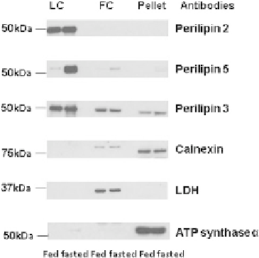
To assess the effectiveness of this method, heart tissues from fed or 12 h fasted mice
were homogenized. To determine the extent of the purity of the LDs obtained, the
cardiac LD fraction was analyzed with an immunoblot with antibodies against pro-
teins reported to be restricted to specific organelles (
Fig. 8.2
). Organelle-specific
markers for cardiac LDs used are the following: the LD-associated protein perilipin
2, member of the perilipin protein family; for cytosolic proteins, lactate dehydroge-
nase; for ER transmembrane protein, calnexin; for mitochondria protein, ATP
synthase
. Due to the difficulty to homogenize heart and its small amount of cardiac
LDs, it is not possible to directly assess the distribution of organelles across other
fractions due to a limitation to resuspend the isolated LDs in a small volume. The
figure provided is indicative that the selected antibodies against protein-specific
a
FIGURE 8.2
Selected protein expression of specific organelle markers in LD, infranatant, and pellet
fractions isolated by differential centrifugation from heart of fed or overnight fastedmice. Each
lane contains equal volume 20
l of sample loaded for perilipin 2 (Progen Biotechnik, cat.
no. GP4), perilipin 5 (Progen Biotechnik, cat. no. GP31), perilipin 3 (Progen Biotechnik,
cat. no. GP36), calnexin (Abcam, cat. no. 191-100), LDH (Cell Signaling Technology, cat. no.
2012), and ATP synthase
m
(MtioSciences, cat. no. MS502) from fed and fasted mice.
One representative experiment is shown.
a