Biology Reference
In-Depth Information
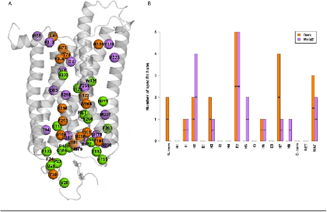
FIGURE 3.4
Comparative hub distribution. In the left panel, hubs are represented as spheres centered on the C
-atoms of the dark rhodopsin structure, on the
C8 atom of retinal, and the O atom of the structural water molecules. Hubs specific to the dark and MII states are, respectively, orange and violet,
whereas those shared by the two forms are green. The contribution of each receptor portion to those hubs that are specific to the dark (orange)
and MII (violet) states is plotted as well in the right panel.
a