Biology Reference
In-Depth Information
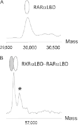
FIGURE 2.4
(A) Positive ESI mass spectra of RARa LBD. (B) Positive ESI mass spectra of RXRa LBD-
RARa LBD. The mass spectra are acquired at V
c
¼
50 V and at a pressure in the interface
of 2.5 mbar. The following molecular weights are measured: 29,930
1.8 Da for RARa
LBD and 56,664
0.3 Da for RXRa LBD-RARa LBD (with deletion of the N-terminal
methionine in RXRa LBD; peak labeled with an asterisk corresponds to species with an
additional N-terminal methionine in RXRa). These results are in agreement with the
molecular weights calculated from the known amino acid sequences.
2.1.3.1
Required materials
- Electrospray time-of-flight mass spectrometer (LCT, Waters)
- Buffer A: 50 mM ammonium acetate pH 6.5
- Purified RXR
a
-RAR
a
LBD heterodimer at 5-10 mg/mL (see the preceding text
for the description of a purification method)
2.1.3.2
Protocol
The instrument is calibrated using the multiply charged ions produced by an injection
of horse heart myoglobin diluted to 2 pmol/mL in a water/acetonitrile mixture (1:1,
v/v) acidified with 1% (v/v) formic acid. Prior to ESI-MS analysis, samples are
desalted on Centricon PM30 microconcentrators (Amicon, Millipore) in buffer
A. Purity and homogeneity of the samples in denaturing conditions are verified
by diluting the complex solution to 5 pmol/mL in a water/acetonitrile mixture
(1:1, v/v) acidified with 1% (v/v) formic acid. Spectra are recorded in the positive
ion mode on the mass range 500-2500 m/z. Verify that the measured molecular
masses are in agreement with those calculated from the amino acid sequences. Sam-
ples are diluted to 10 pmol/mL in buffer A and are continuously infused into the ESI
ion source at a flow rate of 6 mL/min through a Harvard syringe pump.