Biology Reference
In-Depth Information
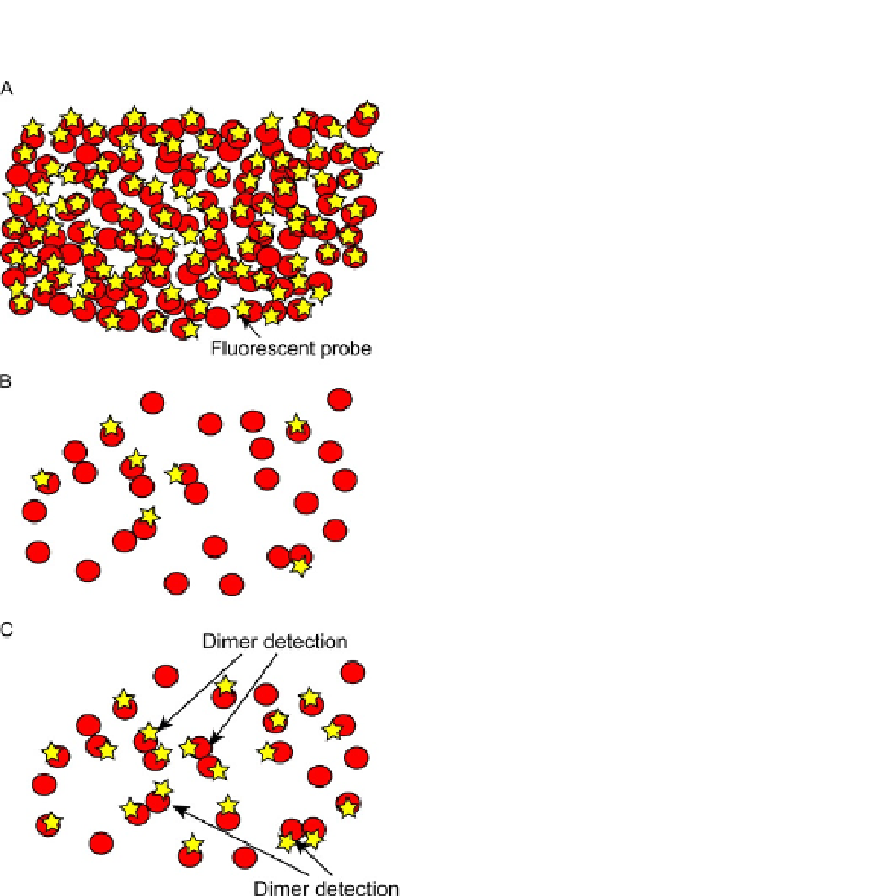
FIGURE 20.2
Receptor expression and fluorescent labeling should be optimized for detection of receptor
dimers and oligomers. (A) Individual fluorescent spots are indistinguishable because
receptors are expressed in cell plasma membranes at a high density (
2 molecules/mm
2
).
(B) Labeling efficiency of receptors with a fluorophore is too low to detect receptor dimers
and oligomers. (C) Low expression density of receptors labeled with fluorophores at
high efficiency is appropriate for detecting receptor dimers and oligomers.
>
(
Medof, Walter, Rutgers, Knowles, & Nussenzweig, 1987
); and 240 copies/
m
m
2
for
folate receptor in MA104 cells (
Rothberg, Ying, Kolhouse, Kamen, & Anderson,
1990
). Therefore, we usually transfect cells with the complementary DNA (cDNA)
of a receptor and express small numbers of a receptor that is not endogenously
expressed in cells, as described in the succeeding text.