Biology Reference
In-Depth Information
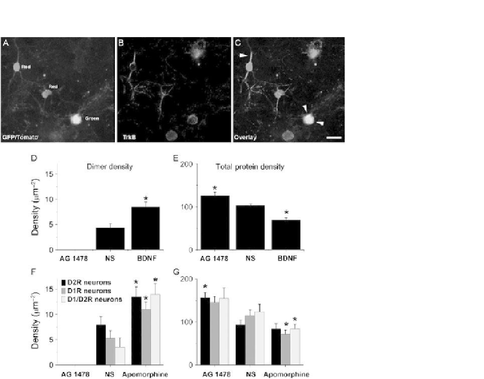
FIGURE 1.2
SpIDA allows the detection and quantification of endogenous TrkB activation by endogenous
dopamine receptors in striatal neurons. (A-C) Surface immunodetection of endogenous
TrkB in striatal neurons prepared from BAC transgenic mice expressing an EGFP (green)
reporter gene in cells having endogenous D2R or a Tomato (red) reporter gene in cells having
endogenous D1R. Arrowheads in C indicate surface TrkB labeling on striatal neurons
expressing different dopamine receptors. Scale bar: 15
m. (D-E) SpIDA analysis of TrkB
dimer (D) and total surface densities (E) of neurons after incubation with AG1478 (0.2 mM for
30 min, n
m
31 neurons/bar), under nonstimulated condition (n
93 neurons/bar), and
¼
¼
following direct stimulation with BDNF (50 pM for 3 min, n
83 neurons/bar). (F-G) SpIDA
analysis of TrkB dimer (F) and total surface densities (G) of neurons after incubation with
AG1478 (0.2 mM for 30 min, n(D2R
¼
¼
30, D1R
¼
33, D1/2R
¼
16) neurons/bar), under
nonstimulated condition (n(D2R
14) neurons/bar), and following
stimulation of endogenous dopamine receptors with apomorphine (2
19, D1R
26, D1/2R
¼
¼
¼
m
M for 3 min,
n(D2R
28) neurons/bar). Data are presented for three
subpopulations of striatal neurons expressing D1R, D2R, or both reporter transgenes. Data
are means
33, D1R
38, D1/2R
¼
¼
¼
SEM.