Biology Reference
In-Depth Information
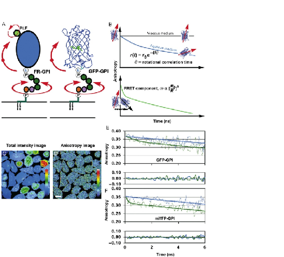
FIGURE 6.1
Time-resolved anisotropy imaging reveals GPI-anchored protein nanoclusters on the
cell membranes. (A) Rotational motions affecting the fluorescent folic acid analog (Pteroyl-
lysyl-folate, PLF) and GFP that label GPI-anchored proteins. (B) Cartoon depicting how the
rotational motions in (A) and homo-FRET can be followed using time-resolved fluorescence
anisotropy. (C-D) Mean fluorescence intensity and anisotropy images of GPI-anchored
proteins on the cell membrane. (E-F) GPI-anchored protein time-resolved fluorescence
anisotropy decays in cholesterol-depleted cells (upper decay lines) and nontreated cells
(lower decay lines). In the presence of cholesterol, GPI-anchored proteins undergo efficient
homo-FRET, resulting in a rapid component in the anisotropy decay (bottom decay lines),
which is absence in cholesterol-depleted cells. This is consistent with a loss in GPI-anchored
protein nanoclustering upon disruption of lipid rafts.
Adapted from
Sharma et al. (2004)
.