Biology Reference
In-Depth Information
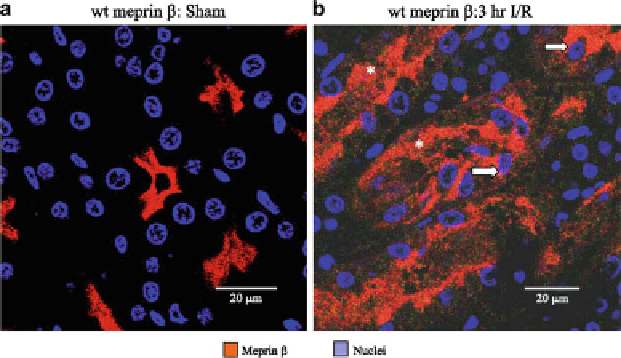
Fig. 4.7 Confocal microscopy of corticomedullary region after 3 h of reperfusion in WT mice.
Kidney sections were immunostained with fluorescent antibody to detect meprin
b
(
red
) and
Hoechst to detect nuclei (
blue
). The image depth is 0.35
m
m. (a) in sham-operated WT mice,
meprin is localized to brush-border membranes extending into the tubular lumen. (b) 3 h after
reperfusion in ischemic mice, meprin localized in luminal regions of the tubules (
asterisks
) and in
the cytoplasm surrounding nuclei of intact cells (
arrows
) (Bylander et al.
2008
)
(Fig.
4.7
). Meprin
KO mice were also less vulnerable to intestinal damage in a
model of experimental IBD induced by dextran sulfate sodium (DSS) (Bond et al.
2006
). Meprin
b
KO mice are more susceptible to severe colitis in the DSS model
(Banerjee et al.
2009
). In contrast, endotoxin (lipopolysaccharide, LPS) induces a
less severe inflammatory response in the
a
KO than the wild-type mice in the model
of urinary tract infection (Yura et al.
2009
). Thus, depending on the type of
challenge, the timing (acute vs. chronic), and the meprin isoform lacking, the
response to a challenge in meprin null mice can be more or less severe than the
wild-type mice.
From the mouse KO investigations, we conclude that meprins act in pericellular
proteolysis in the movement of leukocytes to sites of damage or infection, and in the
cytokine activation/inactivation systemically and/or at site of damage or infection.
For these reasons, we have explored the role of meprins in the hematological system
(leukocytes) and in affecting cytokine catabolism in the in vivo models with the
meprin KO mice.
a
4.5 Meprins Role in the Distribution of Leukocytes
The recently available meprin knockout mice have provided much of the informa-
tion known regarding the function of the enzymes in vivo, and a picture of the
pathological consequences of lacking these enzymes is emerging. Quantitative