Biology Reference
In-Depth Information
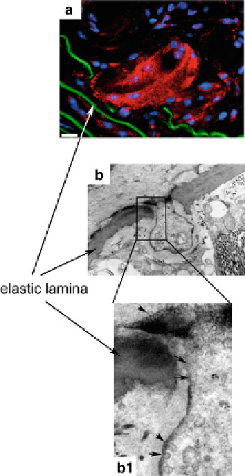
Fig. 2.6 (a) Cathepsin
K-expressing multinucleated
cell at the site of an elastic
lamina break in the
brachiocephalic artery of
Apoe
/
mice (
red
:
cathepsin K staining). (b)
Electron microscopy image of
a cellular podia passing
through an elastic lamina
break. (b1 depicts a higher
magnification of b where a
multitude of vesicles merge
with the outer cell membrane
suggesting the release of
lysosomal cathepsins at the
elastin break site.
Bar
in a
represents 20
m;
magnification for b and b1:
29,000
m
) (modified after
Samokhin et al.
2008
)
2.5.3 Lung (Collagenolytic and Elastolytic Cathepsins)
Lung fibrosis is a pulmonary disorder characterized by ECM deposition, alveolar
epithelial injury, and scar tissue formation. Lung fibroblasts from fibrotic tissue have
increased cathepsin K activity and it is speculated that cathepsin K is needed to fight
the excessive collagen deposits (Buhling et al.
2004a
). Cathepsin K-deficient mice
are more prone to develop bleomycin-induced lung fibrosis. They deposit signifi-
cantly more ECM when compared with wild-type mice, suggesting a role for
cathepsin K in the regulation of lung matrix, likely due to the lack of collagen
degradation in fibroblasts (Buhling et al.
2004a
). More recently, it was also shown
that the overexpression of cathepsin K in a pulmonary fibrosis mouse model reduced
lung collagen deposition and improved lung function (Srivastava et al.
2008
).
In inflammatory conditions such as emphysema the lung ECM is destroyed by
unwanted proteolytic action. Both MMP and cathepsin (B, S, L, H, and K) expres-
sion has been shown to be increased in mouse models of IL-13-stimulated chronic