Agriculture Reference
In-Depth Information
conveyed by the portal vein to the liver. The liver is
drained by the hepatic veins which enter the posterior
vena cava, wherein the blood is conveyed to the heart.
Bacteria or parasites which gain entry to the portal
vein may be arrested within the sinusoids of the liver,
but this organ is an imperfect filter and organisms may
pass through to the heart and thence to the lungs. For
example, hydatid cysts may be found in the lungs and
occasionally immature liver flukes in the lungs of cattle
and older sheep but not in pigs.
the
myocardium
, has a smooth lining, the
endocardium
,
to its four cavities (left ventricle and atrium, right ventri-
cle and atrium). Covering the cardiac muscle is the
epicardium
, the visceral layer of the pericardium.
In reality, the
circulation
consists of two pumps, the
left and right sides, the former being involved with the
systemic circulation and the latter the pulmonary
circulation.
The heart is reddish brown in colour in all the food
animals; the myocardium has a firm consistency, and
the epicardium and endocardium are smooth and glis-
tening. The right and left ventricles may be readily
distinguished by palpation, the wall of the left ventricle
being three times as thick as that of the right, while the
mitral valve and its chordae tendineae are stronger than
the tricuspid valve of the right side. A certain amount of
blood clot is found normally in each of the ventricles
after death.
Spleen (melt)
The spleen is not essential to life. In the foetus, it forms
red and white cells, lymphocytes being produced during
the life of the animal. It also acts as a storage for RBCs
and for the destruction of old red cells and platelets.
Antibodies are formed in the spleen, which, in certain
diseases, for example, anthrax or trypanosomiasis,
becomes very enlarged.
Ox
The ox heart shows three ventricular furrows on its sur-
face. Two
ossa cordis
, which are cartilaginous until 4
weeks after birth, develop at the base of the heart in the
aortic wall. The ox heart weighs 1.8-2.2 kg. In pregnant
cows and in those with a septic infection, it is frequently
pale, flabby and friable.
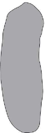
Ox (Fig. 2.11)
The spleen of the ox is related to the left dorsal side of the
rumen and also to the diaphragm. In the young bovine,
it is reddish brown, elongated and slightly convex with
rounded edges; lymph follicles are apparent on the cut
surface. In the cow, the organ is bluish and flat with sharp
edges and rounded extremities; it weighs 0.9-1.3 kg.
Sheep
There are three ventricular furrows, while in later years,
a small
os cordis
may develop on the right side. The heart
weighs 85-113 g.
Dorsal extremity
Pig
Only two ventricular furrows are normally present in the
pig heart although a rudimentary posterior furrow may
be present. The apex is more rounded than in sheep, and
the heart cartilage ossified in older animals. The weight
is 170-198 g.
Splenic vein
Splenic artery
Hilus
1
Horse
The heart has two ventricular furrows, the aortic cartilage
becoming partly ossified in older animals. The average
weight is 2.7 kg, although much greater in racehorses; in
the thoroughbred horse Eclipse, the heart weighs 6.3 kg.
2
3
Portal circulation
The portal circulation is important in the study of the
spread of certain parasitic and bacterial infections
throughout the body.
The portal vein is formed by two main branches, the
gastrosplenic and mesenteric veins which drain the
stomach and intestines. The veins also drain blood
from the pancreas. Venous blood from these organs is
Ventral extremity
Figure 2.11
Spleen of ox, visceral surface. 1, area of attachment
to rumen (non-peritoneal); 2, caudal border; 3, line of peritoneal
reflection.


