Agriculture Reference
In-Depth Information
the remainder of the omasum, the abomasum and the
intestine. The
reticulum
, which is placed transversely
between the anterior extremity of the rumen and the
posterior surface of the diaphragm to which it is adher-
ent, causes a depression on the posterior aspect of the
thin, left lobe of the liver (Fig. 2.2). The omasum and
abomasum are attached to the posterior surface of the
liver by means of the
omentum
or
caul fat
, the root of
this membrane being apparent on the posterior aspect
of the liver to the left of the portal lymph nodes when
the liver is removed from the carcase. The omentum,
after connecting the liver and omasum, is continued
to the lesser curvature of the abomasum and thence
to the duodenum. The anatomical relations of the
bovine stomach play an important part in the aetiology
of traumatic pericarditis. The average capacity of the
stomach is 150 l.
Stomach
Ox (Fig. 2.2, Fig. 2.3 and Fig. 2.4)
The
oesophagus
is comparatively short and wide, meas-
uring about 1 m long and 5 cm wide. The voluntary
muscle, which performs the reverse peristaltic action in
rumination, weighs about 340 g.
The
stomach
(
paunch
) consists of four compart-
ments: the rumen, the reticulum, the omasum and the
abomasum, which is the true digestive stomach and
secretes gastric juice. The
rumen
occupies 75% of the
abdominal cavity; it is bounded on the left side by the
abdominal wall, on the anterior extremity by the reticu-
lum and part of the omasum and on the right side by
Dorsal sac
of rumen
Mucous membranes
Rumen
Brown or black in colour except on pillars or
folds where it is pale and studded with large papillae
Reticulum
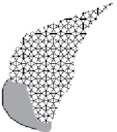
Honeycomb-like appearance with four-, five-
or six-sided cells
Omasum
Prominent longitudinal folds, about 100 in
number, and sometimes called the 'bible'
Abomasum
Some 30 prominent oblique folds in the body
of the abomasum but absent in the pyloric portion
3
1
5
Ventral
sac of
rumen
Omasum
7
2
6
4
Reticulum
Abomasum
A feature of the calf stomach is the relatively large size
of the abomasum as compared with the small size of
the rumen, which remains small until the animal is
weaned. As the calf commences to take solid foods, the
size of the rumen increases until in the adult animal it
represents 80% of the total stomach capacity and the
abomasum 7-8%.
Figure 2.2
Stomach of ox, right side. Oes, oesophagus; 1, insula
between right longitudinal groove below and accessory groove
above; 2, caudal groove of rumen; 3 and 4, right dorsal and ven-
tral coronary grooves; 5 and 6, caudodorsal and caudoventral
blind sacs; 7, pylorus. The positions of the reticulum, omasum and
abomasum have been altered by removal of the stomach from the
abdominal cavity and inflation.
L.K.
Dorsal sac
Sp
Oes.
b.s
ʹ
b.s
ʺ
Ret.
Ventral sac
Figure 2.3
Projection of viscera of cow on body wall, left side. b.s., atrium of rumen; b.s.
′
, b.s.
″
, blind sacs of rumen; O, ovary; Oes,
oesophagus; Ret., reticulum; Sp, spleen. The left kidney (L.K.) is concealed by the dorsal sac of the rumen and is indicated by dotted lines.
The median line of the diaphragm is dotted.


