Agriculture Reference
In-Depth Information
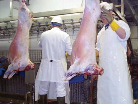
Figure 9.9
Traditional post-mortem inspection of lamb carcases
(Reproduced with permission from Harold Moore).
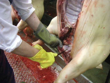
Figure 9.10
Traditional post-mortem inspection of the porcine
head and submaxillary lymph nodes (Reproduced with permission
from Harold Moore).
Skin lesions are an important diagnostic feature of
swine erysipelas, swine fever and urticaria. The skin
should also be examined for 'shotty eruption, the tail for
necrosis, the feet for abscess formation and the udder for
actinomycosis.
The viscera require inspection in the manner detailed
for cattle, with particular attention to pneumonia and
the secondary complications that develop in virus pneu-
monia, mainly pleurisy, pericarditis and, to a lesser
extent, peritonitis.
The submaxillary lymph nodes are routinely examined
for TB. Abscesses in the submaxillary lymph node may be
caused by the passage of sharp foreign bodies through the
wall of the pharynx or, in some countries, a beta-haemo-
lytic
streptococcus
which also often causes tongue
abscesses. Small yellow, necrotic foci resembling TB but
caused by
Corynebacterium equi
are sometimes found in
these nodes. The presence of metal spicules in the dor-
sum of the tongue has been identified as a problem in the
United Kingdom by the manufacturers of pressed tongue.
Some of these fragments have been identified as hypo-
dermic needles, but others are pieces of wire and would
appear to have originated from car tyres given to the pigs
as 'toys. The liver need not be incised except when it
appears cirrhotic. The kidney surface should be exam-
ined for cysts and systemic changes (Fig. 9.10).
Where
Cysticercus cellulosae
is prevalent, the
investigation must include examination of the directly
visible muscular surfaces, in particular the thigh mus-
cles, the pillars of the diaphragm, the intercostal muscles,
the heart, the tongue and the larynx and, if necessary,
the abdominal wall and the psoas muscles freed from
fatty tissue. Where trichinosis is known or suspected,
appropriate examination and muscle sampling must be
carried out.
Current post-mortem inspection in the pig requires
that only the submaxillary lymph nodes and the
supramammary lymph nodes in sows are routinely
incised. Within the EU, the mesenteric lymph nodes of
pigs are no longer incised because of the frequent con-
tamination of knives with
Salmonella
organisms which
may be present in the nodes.
Traditional post-mortem inspection of equines
Post-mortem inspection of equidae follows the same gen-
eral pattern for cattle and all other livestock. Although equi-
dae generally possess fewer lesions than other animals,
particular attention should be paid to the lungs and liver for
evidence of echinococcal cysts and to the muscles and
lymph nodes for melanosis. The main carcase lymph nodes
should be examined when systemic or generalised disease is
suspected, when TB lesions are detected and when the live
animal has shown a reaction to the mallein test. The possi-
bility of glanders requires that the mucous membranes, tra-
chea, larynx, nasal cavities, sinuses and their ramifications
are carefully examined, after splitting the head in the
median plane and excision of the nasal septum.
Traditional post-mortem inspection of poultry
Facilities should be available for whole-carcase inspec-
tion after defeathering and washing. Cases with obvious
disease, fractures, injuries, blood blisters, etc. can be
detected and detained at this stage.
Second-stage inspection takes place on the partially
eviscerated carcase where it is possible to relate car-
case and viscera. The viscera, hock joints and tibias
are observed and the latter palpated. The body cavity
and internal organs are viewed. In some cases,
one inspector examines the carcase, while another