Agriculture Reference
In-Depth Information
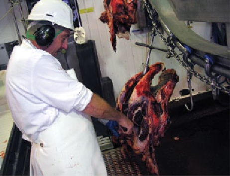
(a)
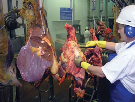
(b)
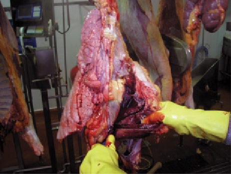
(c)
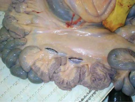
(d)
Figure 9.8
Traditional post-mortem inspection of (a) the bovine head, (b) bovine red offal, (c) bovine heart and (d) bovine mesenteric
lymph nodes (Reproduced with permission from Harold Moore).
The situation with regard to the
gastric
and
mesenteric
lymph nodes
in
cattle
is more problematic. Historically,
examination of TB reactors (1978 in Northern Ireland)
showed that 1.9% of the cattle had lesions in the mesen-
teric lymph nodes only,
Mycobacterium bovis
being
recovered from these lesions. In spite of this finding,
however, it is likely that the saving in time and costs of
inspection outweigh any slight animal health benefits
accruing (Goodhand, 1983).
and main branches of the bronchi opened lengthways,
only when they are to be used for human consumption.
Heart
The pericardium should be examined for evidence of
pericarditis, haemorrhages, etc. The ventricles are then
incised, and the outer and inner surfaces are observed,
particular attention being paid to the presence of petechial
haemorrhages on the epicardium or endocardium and to
cysticerci, hydatid cysts and occasionally linguatulae in
the myocardium. Alternatively, the heart may be everted
after cutting through the interventricular septum with
four lengthways incisions into the septum and the ven-
tricular wall - this latter procedure reduces the heart's
value. A flabby condition of the myocardium is often
associated with septic conditions in the cow, while vege-
tative endocarditis occurs in chronic swine erysipelas and
Lungs
Visual examination, which should be followed by palpa-
tion, should be carried out for evidence of pleurisy, pneu-
monia, TB, fascioliasis, hydatid cysts, etc. The bronchial
and mediastinal lymph nodes should be incised. The lung
substance should be exposed by a long, deep incision
from the base to the apex of each lung, and the trachea