Biomedical Engineering Reference
In-Depth Information
+30 mV
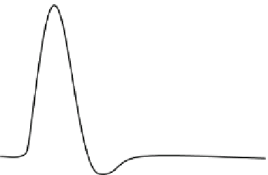
Fig. 2.3
A schematic diagram
showing an action potential,
illustrating electric depolar-
ization and repolarization of
the cell membrane. The mem-
brane potential rises from
−
Repolarization
Depolarization
30 mV within
about 1 ms before repolariza-
tion forces the trend to reverse,
and finally the resting poten-
tial goes back to
−
70 mV
after briefly experiencing a
hyperpolarized state
70 mV to
+
-70 mV
-70 mV
sodium channels start to close. At this repolarization phase, the action potential goes
past the
70 mV level, a state referred to as hyperpolarization. The ion concentration
across the cell gradually returns to the resting level, and the cell returns to the usual
resting potential of
−
70 mV.
As discussed earlier, depolarization across a plasma membrane generates an action
potential. Certain external stimuli reduce the charge across the plasma membrane.
A stimulus may originate from various sources. Mechanical stimuli like stretch-
ing, sound waves, etc. activate mechanically gated sodium channels across the
membrane. Certain neurotransmitters like acetylcholine open ligand-gated sodium
channels. Various electrical impulses may also stimulate and cause depolarization.
The favorable diffusion of sodium ions into the cell locally reduces the membrane's
resting potential. If the reduction is considerable, e.g., if the potential is reduced
to the threshold voltage level (in mammalian neurons, about
−
50 mV), an action
potential is generated in the cell. This kind of action potential usually lasts for less
than 1 ms. Action potentials generated by voltage-gated calcium channels may last
much longer, which is of the order of 100 ms or more. The action potential is very
much organ-specific and is accompanied by various complexities because, in differ-
ent parts of the body, the stimuli appear from different types of sources. For instance,
in some types of neurons, a long burst of rapidly emitted sodium spikes appears due
to the slow calcium spike-induced driving force, whereas, in cardiac muscle cells,
muscle contraction takes place due to the rapid onset of a calcium spike provoked
by an initial fast sodium spike.
−
The Nernst Potential and Membrane Potential
In physiology, the Nernst equation (mentioned above) finds its application in deter-
mining the potential of an ion across a membrane. The general form of the potential
can be written as
RT
zF
ln
[
N
]
out
V
Nernst
=
(2.7)
[
N
]
in

Search WWH ::

Custom Search