Biomedical Engineering Reference
In-Depth Information
0.325
0.33
0.335
10
150000
10
150000
I
10
100000
10
100000
I
10
50000
10
50000
II
10
0
10
0
0.325
0.33
0.335
r
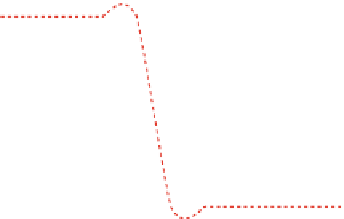
Fig. 5.15
A plot of the energy as a function of the reaction coordinate for gA channels in lipid
bilayer energetics at different orders of screening (single- and double-dashed curves are for the
first- and second-order screening, respectively). I and II represent levels with free energies
G
I
and
G
II
respectively, where gA monomers exist as free (no channel formed) and gA dimer (gA channel
formed).
q
L
electron charge and
other relevant parameters [
1
,
16
,
17
,
35
,
41
,
71
,
75
]) give an estimate of
G
I
and
G
II
to be 10
−
1
and 10
−
8
in first-order and 10
5
and 10
−
4
in second-order lipid screening in units of kJ/mole which
seriously depends on
q
L
as
d
0
increases. The energy orders for
G
I
and
G
II
as mentioned here
are also valid approximations for the corresponding free energy levels presented in the subsequent
Figs.
5.16
,
5.17
,and
5.18
/
q
gA
=
0
.
005,
r
LL
=
7
.
74597 Å.
Ad hoc
assumptions (
q
gA
∼
to compensate to form a stable gA channel which arises mainly from the hydrophobic
binding between the gA channel and the bilayer at the two bilayer channel interfaces.
The smaller the value of
G
I
,
II
, the higher the stability of gA channels. We observe
that the value of
G
I
,
II
for the second-order lipid-screening is orders of magnitude
higher than that for the first-order lipid-screening (higher orders of lipid screening
account for higher values of
d
0
−
l
). Knowing the effective values of charges (in units
of coulombs) on a gramicidin monomer,
q
gA
, and that of a lipid's head group region,
q
L
, one can readily calculate and show in real energy units (J), using the screened
Coulomb interaction theory, that
G
I
has values which are drastically reduced and
hence the value of
l
approaches 0 Å. For exam-
ple, making an
ad hoc
assumption that
q
gA
and
q
L
should be on the order of a few
electron charges, we find
G
I
,
II
collapses as the value of
d
0
−
G
I
,
II
to be on the order of kJ/mole for the first-order lipid
screening, which closely corresponds to the phenomenological bilayer deformation
energy calculated in another study [
13
]. The same
ad hoc
assumption ensures that
G
I
,
II
increases to the order of 105 kJ/mole for the second-order lipid screening.
This drastic increase in bilayer deformation energy requirements for stable chan-
nel formation with increasing the bilayer thickness channel length mismatch causes
gA channel formation to be extremely difficult at a higher order of lipid screening.
Beyond a certain level of hydrophobic bilayer channel mismatch, the deformation
energy reaches values which are outside a biological binding energy scale, which
suggests that at this high energy level the
β
-helical gA channels must experience


Search WWH ::

Custom Search