Information Technology Reference
In-Depth Information
Chapter 5:
MPEG video compression
In this chapter the principles of video compression are explored, leading to descriptions of MPEG-1, MPEG-2 and
MPEG-4. MPEG-1 supports only progressively scanned images, whereas MPEG-2 and MPEG-4 support both
progressive and interlaced scan. MPEG uses the term 'picture' to mean a full-screen image of any kind at one point
on the time axis. This could be a field or a frame in interlaced systems but only a frame in noninterlaced systems.
The terms field and frame will be used only when the distinction is important. MPEG-4 introduces object coding
which can handle entities that may not fill the screen. In MPEG-4 the picture becomes a plane in which one or
more video objects can be displayed, hence the term video object plane (VOP).
5.1 The eye
All imaging signals ultimately excite some response in the eye and the viewer can only describe the result
subjectively. Familiarity with the functioning and limitations of the eye is essential to an understanding of image
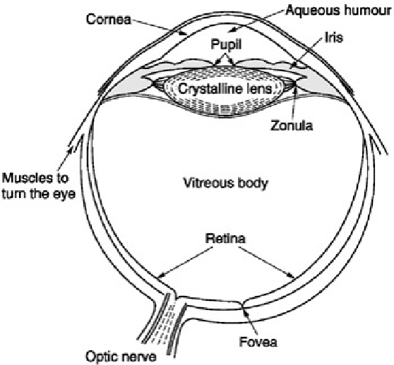
compression. The simple representation of Figure 5.1 shows that the eyeball is nearly spherical and is swivelled by
muscles. The space between the cornea and the lens is filled with transparent fluid known as
aqueous humour
.
The remainder of the eyeball is filled with a transparent jelly known as
vitreous humour
. Light enters the cornea,
and the amount of light admitted is controlled by the pupil in the iris. Light entering is involuntarily focused on the
retina by the lens in a process called
visual accommodation
. The lens is the only part of the eye which is not
nourished by the bloodstream and its centre is technically dead. In a young person the lens is flexible and muscles
distort it to perform the focusing action.
Figure 5.1:
The eyeball is in effect a living camera except for the lens which receives no blood supply and is
technically dead.
In old age the lens loses some flexibility and causes
presbyopia
or limited accommodation. In some people the
length of the eyeball is incorrect resulting in
myopia
(short-sightedness) or
hypermetropia
(long-sightedness). The
cornea should have the same curvature in all meridia, and if this is not the case,
astigmatism
results.
The retina is responsible for light sensing and contains a number of layers. The surface of the retina is covered with
arteries, veins and nerve fibres and light has to penetrate these in order to reach the sensitive layer. This contains
two types of discrete receptors known as
rods
and
cones
from their shape. The distribution and characteristics of
these two receptors are quite different. Rods dominate the periphery of the retina whereas cones dominate a
central area known as the
fovea
outside which their density drops off. Vision using the rods is monochromatic and
has poor resolution but remains effective at very low light levels, whereas the cones provide high resolution and
colour vision but require more light. Figure 5.2 shows how the sensitivity of the retina slowly increases in response
to entering darkness. The first part of the curve is the adaptation of cone or
photopic
vision. This is followed by the
greater adaptation of the rods in
scotopic
vision. At such low light levels the fovea is essentially blind and small
objects which can be seen in the peripheral rod vision disappear when stared at.