Information Technology Reference
In-Depth Information
sound travelling though a fluid. Sound enters the cochlea via a membrane called the oval window. If airborne sound
were to be incident on the oval window directly, the serious impedance mismatch would cause most of the sound to
be reflected. The middle ear remedies that mismatch by providing a mechanical advantage.
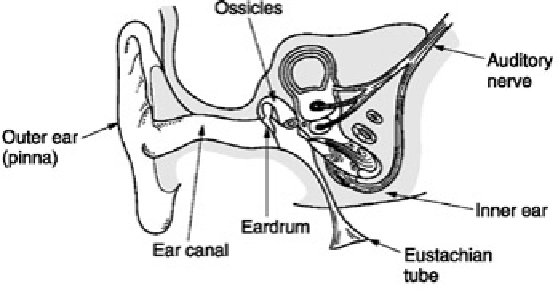
Figure 4.8:
The structure of the human ear. See text for details.
The tympanic membrane is linked to the oval window by three bones known as ossicles which act as a lever
system such that a large displacement of the tympanic membrane results in a smaller displacement of the oval
window but with greater force.
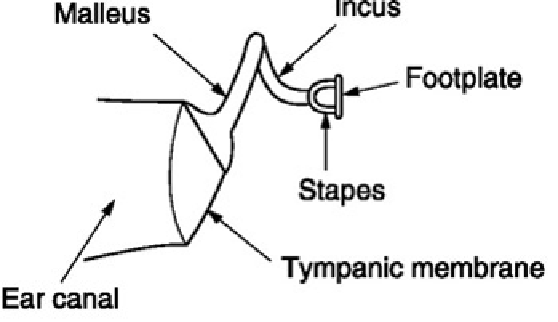
Figure 4.9
shows that the malleus applies a tension to the tympanic membrane
rendering it conical in shape. The malleus and the incus are firmly joined together to form a lever. The incus acts
upon the stapes through a spherical joint. As the area of the tympanic membrane is greater than that of the oval
window, there is a further multiplication of the available force. Consequently small pressures over the large area of
the tympanic membrane are converted to high pressures over the small area of the oval window. The middle ear
evolved to operate at natural sound levels and causes distortion at the high levels which can be generated with
artificial amplification.
Figure 4.9:
The malleus tensions the tympanic membrane into a conical shape The ossicles provide an
impedance-transforming lever system between the tympanic membrane and the oval.
The middle ear is normally sealed, but ambient pressure changes will cause static pressure on the tympanic
membrane which is painful. The pressure is relieved by the Eustachian tube which opens involuntarily whilst
swallowing. Some of the discomfort of the common cold is due to these tubes becoming blocked. The Eustachian
tubes open into the cavities of the head and must normally be closed to avoid one's own speech appearing
deafeningly loud. The ossicles are located by minute muscles which are normally relaxed. However, the middle ear
reflex is an involuntary tightening of the
tensor tympani
and
stapedius
muscles which heavily damp the ability of the
tympanic membrane and the stapes to transmit sound by about 12 dB at frequencies below 1 kHz. The main
function of this reflex is to reduce the audibility of one's own speech. However, loud sounds will also trigger this
reflex which takes some 60-120 milliseconds to operate; too late to protect against transients such as gunfire.
[
3
]
Moore, B.C.,
An Introduction to the Psychology of Hearing
, section 6. 12, London: Academic Press (1989)
4.5 The cochlea
The cochlea is the transducer proper, converting pressure variations in the fluid into nerve impulses. However,
unlike a microphone, the nerve impulses are not an analog of the incoming waveform. Instead the cochlea has
some analysis capability which is combined with a number of mental processes to make a complete analysis. As
shown in
Figure 4.10
(a), the cochlea is a fluid-filled tapering spiral cavity within bony walls. The widest part, near
the oval window, is called the
base
and the distant end is the
apex
.
Figure 4.10
(b) shows that the cochlea is
divided lengthwise into three volumes by Reissner's membrane and the basilar membrane. The
scala vestibuli
and
the
scala tympani
are connected by a small aperture at the apex of the cochlea known as the
helicotrema
.
Vibrations from the stapes are transferred to the oval window and become fluid pressure variations which are
relieved by the flexing of the round window. Effectively the basilar membrane is in series with the fluid motion and