Geography Reference
In-Depth Information
11.2.1 Photons of light turned into information
Neurobiologists have probed deeper within real vision systems to gain a better understanding
of the biological and organizational arrangement of this visual 'belief engine' (Kandel,
Schwartz and Jessell, 1991; Thompson, 2000). Once light is absorbed by the retina it is
translated into neural information, to become a set of synaptic responses. The retina at
the back of the eye consists of about 125 million photoreceptors - one-third are cones
that are used for colour vision and two-thirds are rods that are used for grey-scale vision.
Probabilistically it is possible for a single photon to excite one rhodopsin molecule in a rod,
causing about 500 molecules of transducin to be released, resulting in the blocking of about
1 million sodium ions in the rod that then results in a change of synaptic response (Kingsley,
Gable and Kingsley, 1996). So in the right conditions, that is, a dark room and no stimulus
for 20 plus minutes, a single photon on the retina can be visible - which shows one of the
many ways the evolution of the human visual system is at both the biological and physical
limits. Unfortunately, knowing the limits of the system do not tell us about the way we see
whole objects and explore in our case geographic structures and maps.
Although there are about 125 million receptors, there are only about one million ganglion
cells that are used to transmit this information from the retina to the brain along the optic
nerve. These signals also flow relatively slowly, resulting in a communication bottle-neck
that requires about a 250:1 compression ratio. This means a lot of data will be lost. In the
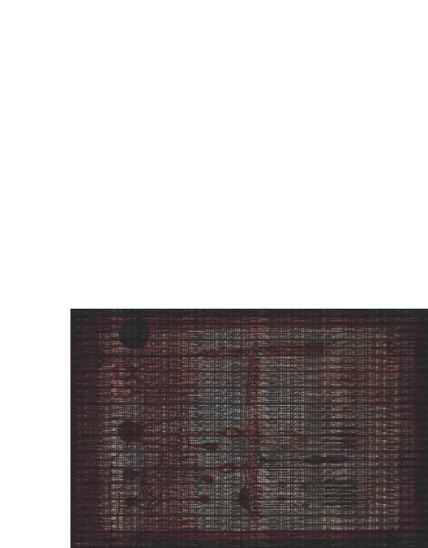
human eye this is achieved by intermediary cells, illustrated in Figure 11.1, that are positioned
between the retina and the optic nerve. These cells consider differences in intensity value
Figure 11.1
Structure of the mammalian retina (source
ca
1900 by Santiago Ramon y Cajal). Light
enters from the left and the rods and cones are shown on the far right. In the middle are the
intermediary cells with connectors going to the optic nerve on the far left


Search WWH ::

Custom Search