Biomedical Engineering Reference
In-Depth Information
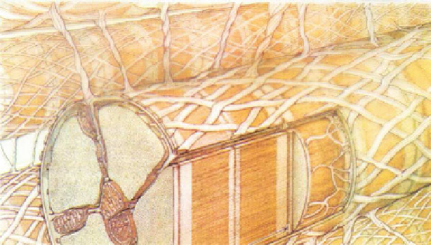
Fig. 5.1
Cardiomyocyte, its collagen envelope and struts; the sarcolemma and its T tubules; inside,
the sarcomere, mitochondria and sarcoplasmic reticulum (from [
331
]).
between cardiomyocytes and between them and perfusing capillaries allow bulk
motions during the cardiac cycle.
Four types of connective tissues can thus be distinguished: (1) an endomysium-
like connective tissue made of beams of collagen fibers that connect cardiomyocytes;
(2) connective tissue that links cardiomyocytes and capillaries; (3) perimysium-
like connective tissue that forms a sheath of spiral collagen fibers around
cardiomyocytes; and (4) epimysium-like connective tissue, i.e., layers of elastin
and collagen fibers that limit the endocardic and epicardic surfaces.
5.2
Internal Organization of Cardiomyocytes
Cardiomyocytes contain structural and functional compartments with the nucleus,
array of myofibrils, sarcoplasmic reticulum, and mitochondria. These densely
packed organelles communicate via functional junctions (e.g., between the sar-
coplasmic reticulum and T tubules for electromechanical coupling) and structural
contacts (e.g., between the sarcoplasmic reticulum and mitochondria for the control
of Ca
2
+
homeostasis).
5.2.1
Sarcolemma
Cardiomyocyte sarcolemma contains many ion channels, pumps, and exchangers
that shape the action potential, the depolarization wave that is received and
transmitted to trigger the contraction. Many receptors for extracellular ligands such
Search WWH ::

Custom Search