Biology Reference
In-Depth Information
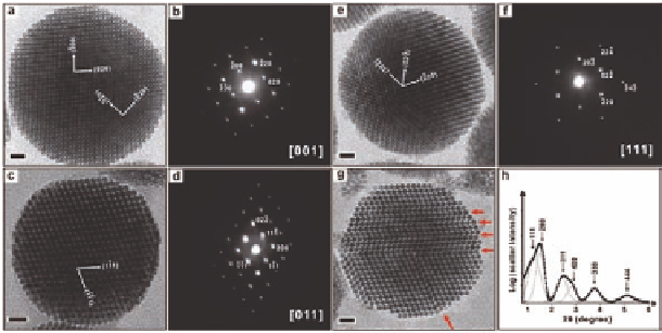
Moreover, the supercrystalline structure of these colloidal
superparticles was determined by further TEM and small-angle
electron diffraction (SAED) studies (Fig. 13.11). TEM clearly shows
that these particles exhibit on-axis superlattice-fringe patterns
that are related to a face-centered cubic (fcc) superlattice structure
with a lattice constant of 11.7
0.2 nm. The [001] image shows the
perpendicular cross-fringes projected from the {200}
±
and {220}
SL
The
cross-fringes in the [011] projection image exhibit an angle of 70.5
planes of the superlattice, respectively (Fig. 13.11a,b).
45
SL
°
(Fig. 13.11c,d), which is consistent with the theoretically expected
value of 70.53
planes. The [111]
projection image shows the characteristic hexagonal cross-fringes
with an interdot spacing of 4.7 nm (Fig. 13.11e,f ), which is much
smaller than the size of the artificial atom building blocks (5.8 nm
Fe
°
between the (1 11)
and (111)
SL
SL
nanocrystals) but precisely related to the spacing of 4.1 nm
between the {022} planes in the superlattice. These results indicate
that these superlattice fringes may not be direct images of the
nanocrystal artificial atoms but provide their superlattice-spacing
information. This fact suggests that the origin of these superlattice
fringes is from electron phase contrast due to the interference
O
3
4
Figure 13.11
(a) TEM image of a superparticle viewed along the [001]
zone axis and (b) the SAED pattern taken from this particle;
(c) TEM image of a superparticle viewed along the [011]
zone axis and (d) the SAED pattern taken from this particle;
(e) TEM image of a superparticle viewed along the [111]
zone axis and (f ) the SAED pattern taken from this particle.
Scale bars: 20 nm. From Ref. [1] with permission.


Search WWH ::

Custom Search