Biology Reference
In-Depth Information
the sample, preferentially bind to the immobilized bio-GBP1 owing
to a specific biotin
−
streptavidin conjugation, thereby allowing for
fluorescence imaging to reveal the areas of immobilization. Figure
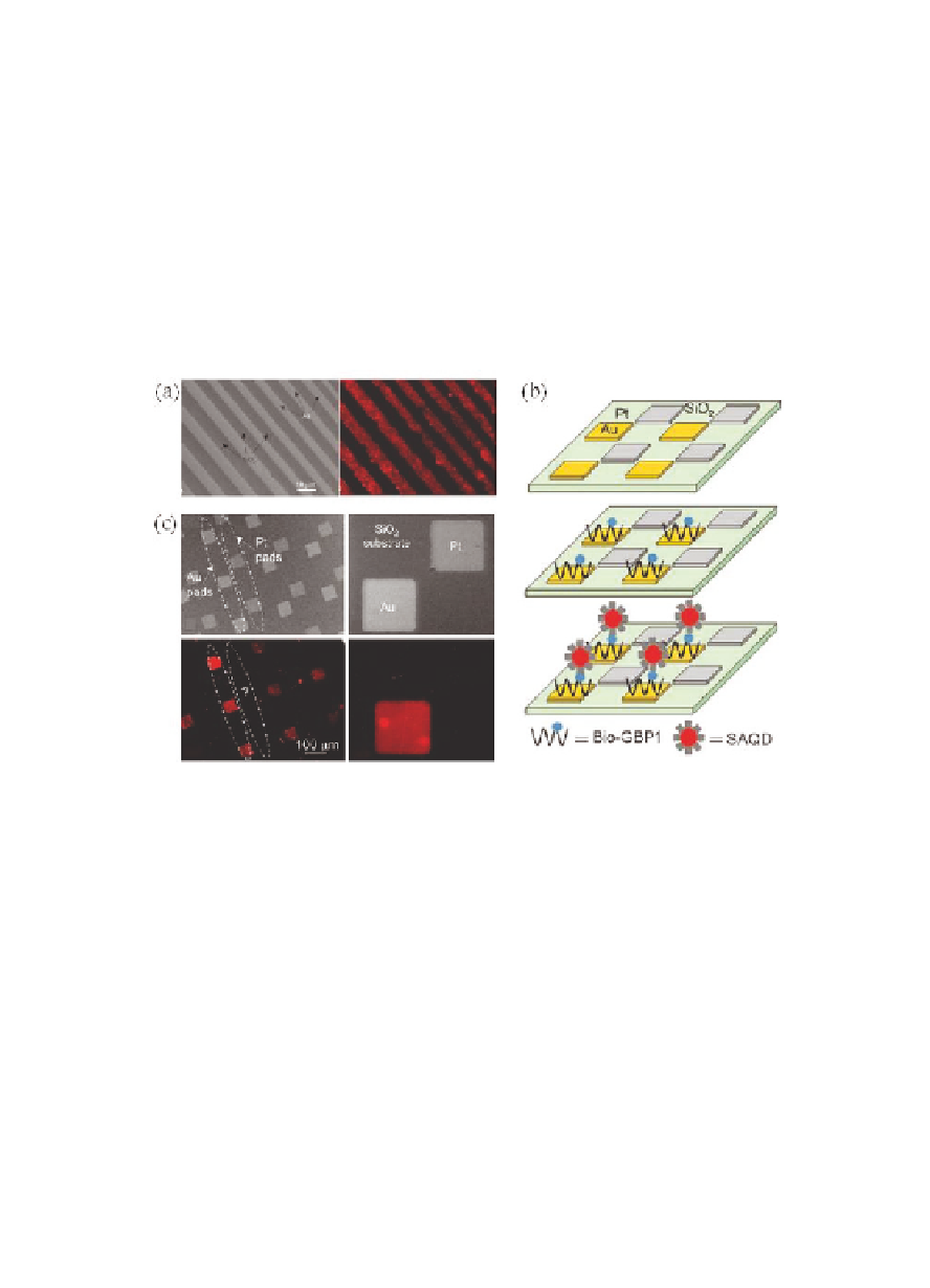
12.2 displays an optical micrograph of the patterned substrate as
well as a fluorescence microscopy image exhibiting defined regions
of SAQDs immobilization on the self-immobilized bio-GBP1. Red-light
emission was recorded from the Au lines whereas no color reversal
developed in the SiO
regions. Hence, the genetically engineered
peptide was concluded to preferentially bind to Au substrates rather
than the oxide surface even in the presence of the biological linker
biotin [1].
2
Figure 12.2
Directed self-assembly of QDs on GBP1 on Au substrates. (a)
Optical micrograph of patterned Au lines on a SiO
substrate
(top). Fluorescence imaging verifies anchoring of SAQDs
onto bio-GBP1 selectively immobilized on Au lines (bottom).
(b) Schematic of fabrication process. Au and Pt squares are
deposited via sputter deposition on a SiO
2
substrate. Bio-
GBP1 binds preferentially to the Au squares and acts as an
anchor for the subsequent selective deposition of QDs. (c)
Optical micrographs of substrate with Pt and Au pads (top).
Fluorescence images of SAQDs selectively anchored to Au
substrates only (bottom). Reproduced with permission from
Ref. [1]. Copyright Wiley-VCH Verlag GmbH & Co. KGaA.
2
Another
experiment
involved
sputter
deposition
of
the
two noble metals as alternating squares on a SiO
substrate to
2
Search WWH ::

Custom Search