Biology Reference
In-Depth Information
ether to a concentrated solution of
typically produces a stable gelly
material, which small-angle X-ray diffraction (SAXS) demonstrated
to be an unusual lyotropic liquid crystalline phase [24]. CD spectra,
recorded in chloroform at different temperatures and concentrations,
showed weak signals, without any exciton pattern, suggesting that
no supramolecular chirality was originated by self-assembly of
guanosines in these conditions.
1
1
show broad
signals if compared to the ones recorded in the strongly competing
solvent DMSO-d6, as expected for largely associated molecules.
The existence of oligomeric structures in CDCl
H-NMR spectra in CDCl
3
was supported by
ESI mass spectrometry [25]. Moreover, when increasing guanosine
concentration (or lowering temperature), progressive deshielding
of both the imino N
3
H protons takes place, indicating
that the H-bond donor groups of the guanine bases are progressively
involved in the self-assembly process. IR spectra lead to the same
conclusion.
Hence, the lack of added cations and all of the spectral data ruled
out the presence of stacked G-quartet aggregates: the structural
elements giving rise to the mesophase had to be the result of a
different form of LG self-assembly. The structure of these aggregates
was characterized in the solid state (by X-ray diffraction MAS-NMR
spectroscopy and AFM), in solution (by NMR spectroscopy and ESI-
MS) and at graphite-solution interface (by STM) (Fig. 4.11) [25].
1
H and amino N
2
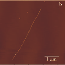
Figure 4.11
Left: current STM survey image of self-assembled architecture
of LG recorded at the solid-liquid interface on HOPG in the
absence of added cations. Right: SFM on mica reveals a single
dry nanoribbon of LG on mica (up to 8 mm long).
Two different ribbon-like aggregates, with different patterns of
hydrogen bonds, were identified in the solid state and in chloroform
solution. The first species (ribbon A, Fig. 4.12A), which is stable in


Search WWH ::

Custom Search