Biomedical Engineering Reference
In-Depth Information
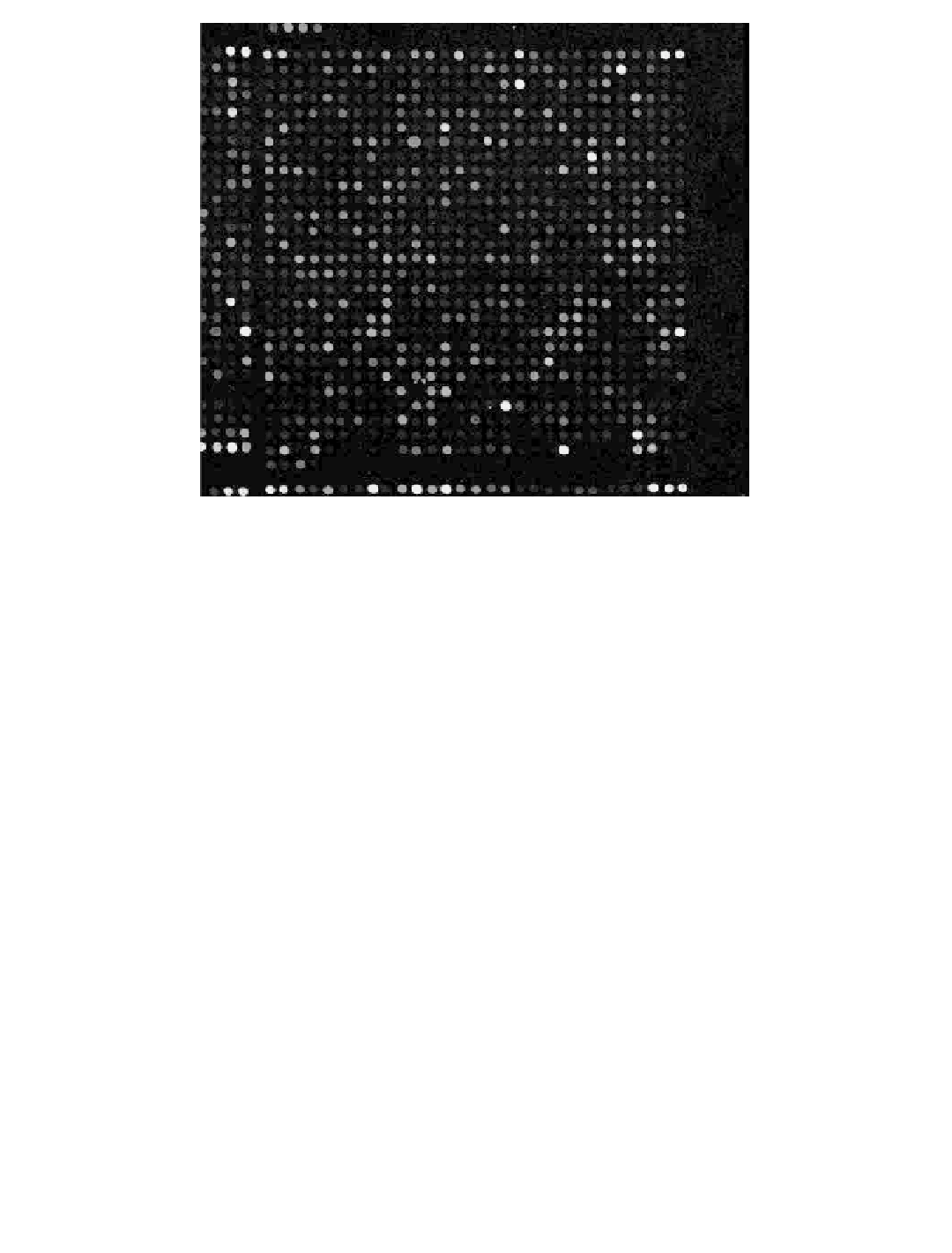
FIGURE 1.51
(See color insert)
A low-resolution fluorescence light microscope image of the spotted array surface of a typical
microarray experiment. Reprinted from website http://www.microarray.org/sfgf/jsp/home.jsp (accessed May
27, 2005). with permission of website.
data analysis company cofounded by the author, that specialized in the application of inte-
grated high-dimensional machine learning and visualization techniques to partners' data sets
in the biotechnology and pharmaceutical industry. Working collaboratively with AnVil, we
applied these data mining techniques to the following two biosensor systems as examples.
The first application was to the microarray biosensor analysis of mRNAs from known cancer
tissue samples to create a cancer tissue classifier for undiagnosed clinical samples. The sec-
ond application was to the analysis of whole cell biosensor output from the cellular testing of
tens of thousands of compounds for cancer efficacy in the National Cancer Institute's (NCI)
repository, to create compound subsets that target-specific cancer cell types.
In the first biosensor case, the object was to create a diagnostic three-class classifier
using a supervised machine learning approach that could distinguish between normal
lung tissue and two subclasses of nonsmall cell lung carcinoma (non-SCLC)—adenocarci-
noma and squamous cell carcinoma. The classifier would be based upon the results of
nucleic acid microarray biosensor experiments testing the mRNA populations from a
number of each of these three clinical tissue types diagnosed via traditional pathological
criteria (176,177). A total of 75 patient samples were studied, consisting of 17 normals, 30
adenocarcinomas, and 28 squamous cell carcinomas. A second data set used was in the
public domain and consisted of 157 samples, including 17 normals, 139 adenocarcinomas,
and 21 squamous cell carcinomas (178). The data were obtained using Chip A from the
Affymetrix Human Genome U95 Chip Set (179). Chip A is a microarray containing spot-
ted sequences for 12,000 full-length genes as well as controls. In a preliminary two-class
(normal lung vs. non-SCLC tissue) examination of the data, the measured microarray
biosensor levels from ~2000 genes were found to be statistically significant (
p
0.05) in
differentiating clinical samples between these two tissue classes. For the more interesting