Biomedical Engineering Reference
In-Depth Information
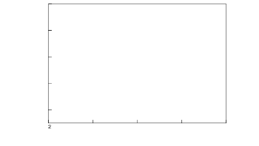
to a limiting value that changed nearly 100% of the original magnitude at the highest doses
used. The response occurred in a dose-dependent fashion, over a 5-6-h period following
drug addition. This effect is consistent with nocodazole's disruption of microtubules. By
contrast, using this biosensor with a 10
M taxol addition, an FDA-approved and widely
used cancer chemotherapeutic drug, caused little initial time-dependent alteration in fre-
quency, consistent with it's known microtubule hyperstabilization effect (98,99). When we
plotted the total biosensor frequency decreases vs. the increasing nocodazole concentration
added, as shown in Figure 1.35, we observed a dose-response curve. This sigmoid curve
shape fit to the data is typical for small-molecule drugs interacting with their biological tar-
gets. The midpoint of this transition is around 900 nM. We corroborated that our cell QCM
biosensor was yielding accurate biological effects by examining the nocodazole range of
this dose curve with fluorescence light microscopy to visualize cellular actin using a rho-
damine-labeled phalloidin chromophore acting as an actin stain. Actin is a known cell
stress marker for surface detachment in these ECs. The range of moderate to progressively
more severe rhodamine-phalloidin staining alterations in cellular actin fibers that we
observed mimicked the range of nocodazole concentrations spanning the biosensor dose
curve. Also in good agreement with the dose curve are nocodazole's cytological effects
observed by light microscopy in various cell types in a number of other studies (97,100).
When the cell QCM biosensor frequency shifts were presented normalized by the number
of cells found firmly attached to the QCM surface following trypsin removal and electronic
counting, then the dose curve did not fall off at the highest concentrations (not shown in
Fig. 1.35). In this case, the curve midpoint was shifted to lower nocodazole concentrations,
indicating that this additional trypsin/cell count measurement creates a more sensitive
drug biosensor.
400
300
200
100
0
0
1
2
FIGURE 1.35
Dose curve of -
f
shift values produced have been
plotted for different experiments carried out at the indicated nocodazole concentrations. A three-component
sigmoid curve has been fit to these data where
R
2
= 0.91. Reprinted from Marx, K.A., Zhou, T., Montrone, A.,
Schulze, H., Braunhut, S. J. (2001). A Quartz Crystal Microbalance Cell Biosensor: Detection of Microtubule
Alterations in Living Cells at nM Nocodazole Concentrations.
Biosen. Bioelectron.
16:773-782. With permission
from Elsevier Publishing.
(
f
) vs. log[Nocodazole] concentration where the maximum