Biomedical Engineering Reference
In-Depth Information
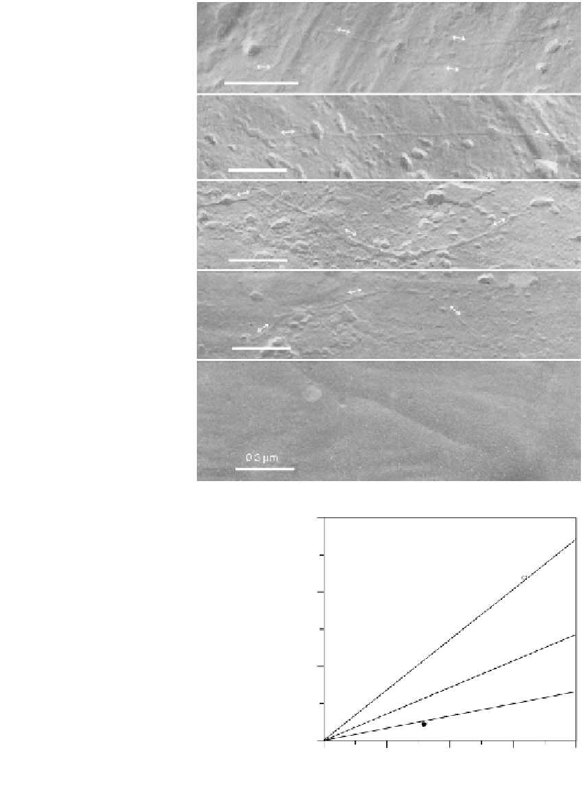
(a)
(b)
FIGURE 1.20
Transmission electron micrographs
from freeze fracture preparations of
linear pBR 322 plasmid DNAs
bound to electropolymerized
polypyrrole films. Panels (a)-(d)
show examples where the entire
length of individual pBR 322 plas-
mid DNAs on the polypyrrole sur-
face is visible, highlighted by the
arrows shown over portions of
their path. Panel (e) is a control
sample of the polypyrrole surface
without DNA bound. The magnifi-
cation bar in all panels indicates 0.3
(c)
(d)
m length. Reprinted from Pande,
R., Ruben, G.C., Lim, J.O., Tripathy,
S., Marx, K.A. (1998). DNA Bound
to Polypyrrole Films: High
Resolution Imaging, DNA Binding
Kinetics and Internal Migration.
Biomaterials
19:1657-1667. With
permission of Elsevier Publishing.
(e)
30
20
FIGURE 1.21
DNA adsorption plotted as a function of
t
0.5
onto
electropolymerized polypyrrole films performed
in 1 mM EDTA, 10 mM Tris, pH 8.0. Three DNA
concentrations were studied: (diamonds) 0.4
10
g/mL, (triangles) 0.2
g/mL, and (circles) 0.1
g/mL. Reprinted with permission from
Minehan, D.S., Marx, K.A., Tripathy, S.K. (1994).
Kinetics of DNA Binding to Electrically
Conducting Polypyrrole Films.
Macromolecules
27:777-783. Copyright (1994) American Chemical
Society.
0
0
2
4
6
8
t
1/2
(min
−
1
)
1.2.2.2 Enzyme Electrode Biosensor—Enzyme Entrapment During
Electropolymerization of Thin Phenolic Films for Hydrogen Peroxide Biosensing
The electropolymerization of thin films using phenolic-based monomers has a number of
attractive features. Phenolic monomers tend to form self-limiting polymer thin films on the
electrode surface that don't exceed thicknesses of about 100 nm (66). This is due to the