Biomedical Engineering Reference
In-Depth Information
3
3
2
2
2
1
1
Membrane current
Light intensity
On
Off
V
c
V
c
V
′
c
0
0
(A)
Clamping voltage
10 pA
1 min
I
p
Membrane current
Light intensity
On
Off
On
Off
Clamping voltage
V
c
= 0 mV
V
c
= 0 mV
V
′
c
=
−
19 mV
(B)
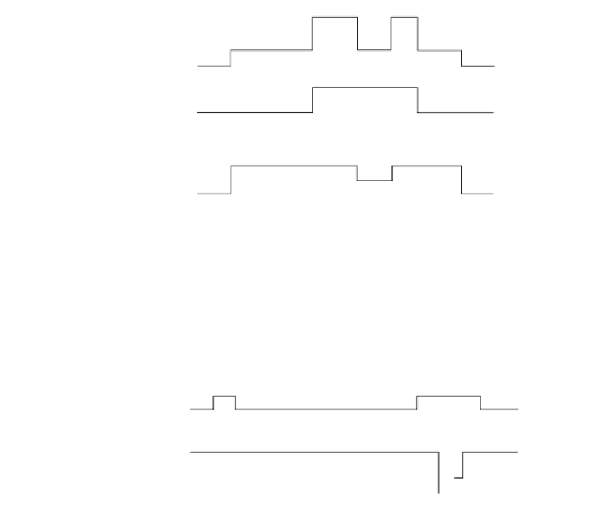
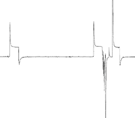
FIGURE 15.12
Null-current method (A, schematic diagram; B, actual experimental record taken from a reconstituted bR-con-
taining BLM). The experimental conditions are as follows: The pH was 6.9, and the temperature was 27.5ºC. The
instrumental time constant was 1 s. The first measurement was a “light on” and “light off” operation, which
allowed
I
p
to be measured (corresponding to the clamping potential 0 mV). In the second measurement, the
clamping potential was adjusted to
19 mV so as to obtain the null-current condition. See the cited source for
detailed experimental conditions. (From Fuller, B. E., Okajima, T. L., Hong, F. T. (1995). Analysis of the d.c. pho-
toelectric signal from model bacteriorhodopsin membranes: d.c. photoconductivity determination by means of
the null current method and the effect of proton ionophores.
Bioelectrochem. Bioenerg.
37:109-124.)
applying an offset voltage,
V
c
), it is sufficient to abolish the DC photocurrent
and move the measured current from level 3 back to level 2 (the preillumination level).
By invoking the principle of potentiometry, the offset voltage can be taken as equal to
the photoemf at the clamping voltage
V
c
, but with an opposite polarity. Thus, the
photoemf is
E
0
(
V
c
E
V
V
E
(15.9)
p
c
c
0
Subsequently, the clamping voltage is then brought back to
V
c
, so as to ascertain that the
measured current actually returns to level 3, which is the level attained before applying
the offset voltage. When the light is finally turned off, the measured current settles back
to level 2, again after a brief AC photoelectric current transient. Finally, with the light