Biomedical Engineering Reference
In-Depth Information
Microscope
objective
Electric field
strength
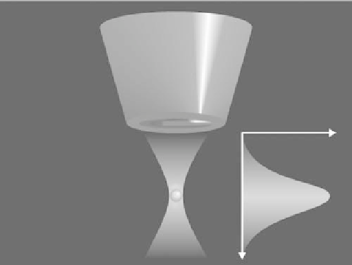
FIGURE 2.2
The principle of “optical tweezers”. A
high-intensity optical beam can be
shaped to form a physical zone that has
variation of electric field intensity. The
gradient of electric field intensity can be
used to trap particles.
Optically trapped
particle
interest. In cases where substantial areas must be interrogated it may be possible to use wide-
field epi-illlumination as an alternative method for single molecule detection.
2.3.4
Wide-Field Epi-Illumination Microscopy
A conventional mode of fluorescence excitation in wide-field microscopy is epi-illumina-
tion, which is perhaps the most straightforward way to image single molecules at the dif-
fraction limit. The optical excitation system consists of a laser source, defocusing optics, a
high-performance dichroic beamsplitter, and an oil-immersion objective with low autoflu-
orescence, where the excitation light and emission light travel through the same optics
(11,37,47). Such instruments often use a multichannel charge-coupled device (CCD) cam-
era or an intensified CCD (ICCD) as a detector. The imaged area is typically about 100
100 µm, but it also depends on the illumination field and the CCD-active area.
This technique has been used to achieve single molecule detection for numerous different
applications. Single myosin molecules that are labeled with one or two copies of a fluores-
cent dye have been studied using this method (21). In this experiment, the large myosin mol-
ecules are adsorbed spontaneously onto a glass slide surface. Schmidt et al. (3) have
measured the random two-dimensional motion of single fluorophore-labeled lipid mole-
cules, which moved slowly in a lipid membrane. These examples indicate that wide-field
epi-illumination can be used to investigate dry surfaces or targets in aqueous media. The use
of a sensitive Peltier-cooled video-rate CCD camera has provided defocused images of sin-
gle molecules so that orientation was observed (48). A comparison of experiment results
with theoretical dipole orientation calculations was accomplished, which used a series of
five images taken with increasing defocusing values ranging from 0 to 1.2 mm in steps of
300 nm. A significant correlation between the fluorescence from images and the patterns that
were calculated for a parallel dipole orientation was seen (48). Owing to the exposure time
of 3 s, only a few molecules survived all exposures without photobleaching or blinking.
The method of wide-field epi-illumination can reduce the amount of time to accumulate
a statistically significant number of observations, but there are technical limitations. While
single molecules are detected when they are in focus, there is often overwhelming back-
ground interference generated from both in-focus and out-of-focus excitation volumes.
The reduction of some background scatter can be achieved by the use of high-quality
optics. Excitation can also be separated from emission radiation by appropriate design of