Biomedical Engineering Reference
In-Depth Information
NSOM fibre tip
Near field excitation
Near field emision
Near field region
(ca. 200 nm)
Fluorophore
Far field
region
Far field excitation
Far field emision
Lens
Confocal
aperture
or pinhole
To detector
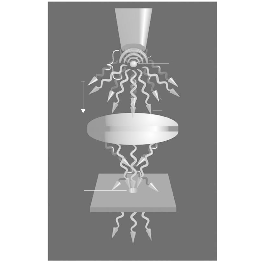
FIGURE 2.1
Physical principles that provide the basis for a number of different single molecule detection methods. An NSOM
tip based on a tapered optical fiber that terminates with a diameter less than that of the excitation radiation is
surrounded by a metal cladding. An evanescent field (wave) surrounds the fiber tip, decaying with distance in
the “near-field” region. A fluorescent molecule (fluorophore) within the near-field region is excited into emission
by the evanescent field, and the emitted radiation scatters into the environment. The emitted radiation is a pho-
ton in the “far-field”, and can be collected by a suitable microscope. A lens serves to focus the emitted light onto
a suitable photodetector. If a pin hole is placed before the detector, it is possible to adjust the depth of focus (focal
plane depth) by restricting the angles of radiation that reach the detector, and this represents the principle of
operation of a confocal microscope.
conformational alterations of these fluorescent molecules (15). By temporally autocorre-
lating the fluorescence intensity, the signal fluctuations can be quantified in terms of
strength and duration. For a small number of molecules present in the detection volume,
the fluctuations in the light intensity are large considering the amplitude and decay rate.
FCS has been used as a tool to study single molecule behavior where the sample volume
is on the order of nano- or pico-liters (29). Very low focal volume and a high quantum
yield of the fluorophore are desired (29).
Rigler and Mets (30) were one of the first teams to use FCS to study single molecules. FCS
has been applied to problems such as determination of rate constants of chemical reactions,