Biology Reference
In-Depth Information
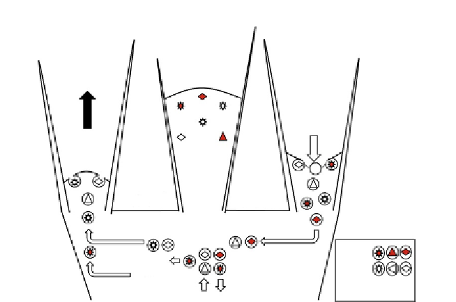
A. Exocytosis
B. Protrusion
C. Endocytosis
Filopodia
Membrane
recycling
Lamellar
initiation
Peripheral
domain
Recycled
New
Legend
D. Sorting
?
Active
Rab5
Local
signaling
Central
domain
Rab7
Inactive
Positive
feedback
From To
Soma
E. Transport
Figure 2.2 Schematic of Trk receptor dynamics during lamellar protrusion and
membrane recycling between the growth cone's central and peripheral domains.
(A) Exocytosis. During lamellar initiation and protrusion (black arrow) in the peripheral
domain, vesicles trafficked from the central domain exocytose, locally adding their
various lipid and protein cargos, including Trk receptors, cell adhesion molecules
(CAM), and other non-Trk receptors (NTR) to the plasma membrane. (B) Protrusion.
As lamellipodia and filopodia (not shown for clarity) protrude outward, Trk receptors
are carried peripherally into the extracellular environment. (C) Endocytosis. Both acti-
vated (red) and inactive (open) receptors and CAMs are returned (open arrow) to the
central domain, endocytosed, and then (D) sorted into distinct endosomes likely based
on their activation state and association with trafficking proteins including the Rab
GTPases. Rab5 and Rab7 appear to direct retrograde (E) transport to the soma for
long distance signaling or degradation, respectively. Rab11 recycling endosomes cycle
inactive receptors back to the plasma membrane for redistribution to peripheral
protrusions. Importantly, activated receptors may also be sorted into local signaling
endosomes that feedback positively to locally perpetuate protrusive activity regionally
within the growth cone. This local signaling may also influence the trafficking of new
vesicular cargoes from the soma, increasing receptor density, and further amplifying
local signaling in growth cone subdomains.
thus bias the direction of central domain advance and the location for future
rounds of exocytosis, protrusion, endocytosis, and sorting. As the previous
central domain consolidates into nascent neurite (
Dent & Gertler, 2003
),
CAMs are endocytosed into vesicles at the base of the growth cone and either
degraded or recycled into new lamellar and filopodial protrusions
(
Kamiguchi & Lemmon, 2000a
) (e.g.,
Fig. 2.1C
). Stabilized protrusions