Biology Reference
In-Depth Information
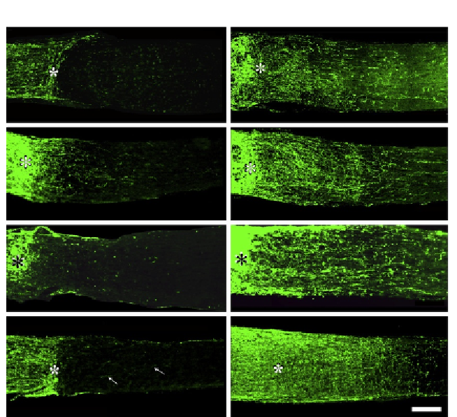
A
B
C
D
E
F
G
H
Figure 6.1 Intraocular inflammation and oncomodulin (Ocm) promote optic nerve re-
generation. GAP-43 immunostaining shows regenerating axons in longitudinal sections
through the mature rat optic nerve 2 weeks after injury. (A, B) Effects of intraocular in-
flammation. Almost no axons grow beyond the site of injury (asterisk) under normal
conditions (A); intraocular inflammation, induced either by lens injury or zymosan injec-
tions, promotes regeneration (B). (C, D) Gain-of-function experiments. Injection of blank
beads into the eye induces slight regeneration that is related to a low level of inflam-
mation (C), whereas beads that release Ocm plus a cAMP analog induce strong regen-
eration (D). (E
-
H) Loss-of-function experiments. Injection of peptide P1 competes with
Ocm for receptor occupancy and suppresses inflammation-induced regeneration (E),
while control peptide P3 has no effect (F). RGCs transfection with ADP ribosyltransferase
C3, which inactivates Rho A, stimulates axon regeneration at a very low level (G, white
arrows). However, transfecting RGCs with the C3 gene and inducing inflammation
strongly enhances regeneration. Scale bar: 200
m
m.
the optic nerve enhances RGCs survival after optic nerve damage, only
the former promotes regeneration (
Ahmed et al., 2010
). Further evidence
for this dissociation comes from studies showing that overexpression
of the antiapoptotic protein Bcl-2 promotes the survival of RGCs after optic
nerve damage without affecting regeneration (
Chierzi, Strettoi, Cenni, &