Information Technology Reference
In-Depth Information
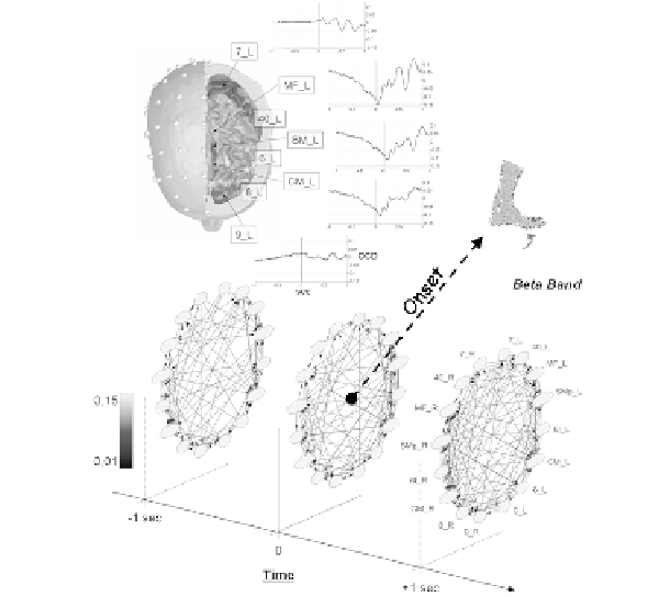
Fig. 10.7. (Up) Realistic head model for a representative subject and cortical activity for the ROIs
in the left hemisphere. (Bottom) Three-dimensional representation of the estimated time-varying
network in the Beta band for the same subject.
centered it on the onset detected by a tibial EMG. The use of the time-varying
Partial Directed Coherence (PDC, see paragraph X.2) to the cortical waveforms
obtained from EEG signals returned a cortical network for each selected time
sample and frequency band. In order to consider only those task-related
connections, a filtering procedure based on statistical validation was adopted. In
each trial, a rest period of 2 seconds preceding the movement was selected as an
element of contrast (from -4 to -2 s before the onset, i.e. the moment in which the
movement occurs). Figure 10.7 illustrates the locations of the regions of interest
(ROIs) on the left hemisphere of the cortex model together with their estimated
temporal activity. At the bottom, the time-varying cortical network in the Beta
frequency band is shown for a representative subject. In particular, three instants
are highlighted; one second before the onset, the onset itself and one second after
the onset.
Figure 10.8a) shows the in-strength values for the average network during
three moments of interest that presented significant differences from random
networks. Among all the cortical regions, the supplementary motor areas of both