Environmental Engineering Reference
In-Depth Information
mesenchymal neoplasms while short ibers did not cause mesothelioma. It is worth noting that
∼8 μm is the maximal size of pore-like structures called stomata on the parietal pleura, the main
route of removal from the plural space. The use of several forms of mineral ibers of differing
physicochemical properties also enabled Stanton to conclude that carcinogenicity of the ibers was
dependent on dimension and durability rather than on physicochemical properties. The importance
of this study and its relevance to future iber toxicology and the setting of iber counting standards
such as that used by the WHO and Occupational Safety & Health Administration (OSHA), cannot
be overstated. Stanton, however, has not been the only one to demonstrate the importance of iber
length, shown in subsequent years in various experimental systems (Davis et al., 1986; Adamson
and Bowden, 1987a,b; Davis and Jones, 1988; Donaldson et al., 1989; Goodglick and Kane, 1990).
The basis of the Stanton hypothesis most likely relates to the role iber length plays in clearance
from its site of deposition. The airways are constructed in such a way that it prevents larger particles
from reaching the lung altogether or reaching beyond the larger, conducting airways. Long ibers
or particles depositing in these larger airways are removed rapidly from the lung via the action of
constantly beating hair-like cilia lining these airways (the mucociliary escalator), which propel
particles and mucus out of the lung. Small particles that can negotiate the airways and deposit in
the proximal alveolar region are phagocytosed with ease by alveolar macrophages, either persist-
ing within the macrophage if durable or moved up onto the mucociliary escalator and ejected from
the lung. Fibers depositing in these non-ciliated regions, due to the aforementioned unusual aero-
dynamic behavior, must be dealt with by alveolar macrophages. The issue of impaired clearance
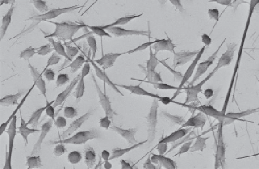
occurs when a iber is longer than the maximal length that a macrophage can comfortably enclose
(∼15 to 20 μm). In this situation, the macrophage will try unsuccessfully to phagocytose the iber
and become “frustrated” as shown in Figure 10.4.
The action of
frustrated phagocytosis
leads to the release of various toxic components, which
are crucial to the normal microbiocidal activity of the cells as part of the innate immune defense,
which can damage surrounding cells and cause inlammation. Inlammation is an important process
and the body's inlammatory cells are critical in maintaining a healthy biological environment.
However, these same protective mechanisms can become damaging, possibly leading to further
damage and disease, when inappropriately activated, uncontrolled, or occurring repeatedly.
If the deposited ibers cannot be cleared via the normal macrophage-mediated means, they may
persist in the alveoli or may translocate out of the lung into the cavity that surrounds the lung (the
pleural cavity), where they may be retained and drive a pathogenic response.
2
FIGURE 10.4
Frustrated phagocytosis. SEM image of mouse alveolar macrophages trying to phagocytose
ibrous material and forming the typical elongated morphology of a frustrated macrophage. (Image copyright
SAFENANO, Edinburgh, U.K.)
Search WWH ::

Custom Search