Biology Reference
In-Depth Information
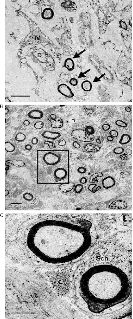
Figure 3.3 Electron microscopic photographs of control (A) and shock wave-treated
peripheral nerves 3 weeks after surgery. Panel (A) shows several degenerated myelin
sheaths (D) engulfed by macrophages (M). A few myelinated regenerated axons
(arrows) too can be seen. In panel (B), a high number of myelinated axons are present