Biology Reference
In-Depth Information
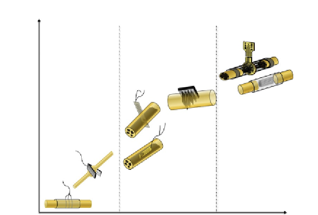
Extraneural
Intraneural
Regenerative
F
E
G
D
C
B
A
Invasiveness/nerve damage
Figure 2.1 Electrodes used to interface peripheral nerves classified according to their
invasiveness and selectivity. Images show examples of (A) cuff electrode, (B) flat inter-
face nerve electrode (FINE), (C) longitudinal intrafascicular electrode (LIFE), (D) trans-
verse intrafascicular multichannel electrode (TIME), (E) multielectrode array (USEA),
(F) sieve electrode, and (G) microchannel electrode.
lumen of the tube (
Hoffer & Loeb, 1980
). Cuff electrodes allow for precise
positioning and significantly reduce stimulus intensity compared to surface
and epimysial electrodes (
Loeb & Peck, 1996
), since the cuff-insulating
sheath limits current leak out of the cuff-nerve space. Compared to
intraneural electrodes, the surrounding approach of cuff electrodes reduces
their selectivity, making them able to record and stimulate only sensorimo-
tor large myelinated fibers and predominantly those located at superficial
locations (
Badia, Boretius, Andreu, et al., 2011
). On the other hand, the
reduced invasiveness of these electrodes makes them easier to handle and
safer to implant (
Naples, Mortimer, & Yuen, 1990
). In order to improve
cuff's selectivity and performance, several strategies, such as multisite cuff
electrodes (
Navarro, Valderrama, Stieglitz, & Sch¨ ttler, 2001; Tarler &
Mortimer, 2004; Veraart,Grill, &Mortimer, 1993;Walter et al., 1997
), inno-
vative cuff structures (
Tyler &Durand, 1997
), complex modes of stimulation
(
Grill & Mortimer, 1996; Navarro et al., 2001
), and advanced processing
algorithms (
Raspopovic, Carpaneto, Udina, Navarro, & Micera, 2010;