Information Technology Reference
In-Depth Information
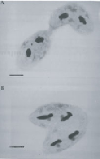
Figure 9.2
(a) A pair of stichotrichs in an early stage
of mating (
Sterkiella histriomuscorum
). The two cells
are connected by a cytoplasmic bridge. (b) A later
stage of mating, in which the two cells are tightly
joined. Bars
=
10
µ
m.
(Figure 9.3a, b). Exchange of DNA now occurs by migration of a haploid
micronucleus from each cell through the cytoplasmic channel into its partner
(Figure 3c). The migratory haploid micronucleus fuses with a resident haploid
micronucleus, forming a new diploid micronucleus in each cell (Figure 3d). The
cytoplasmic channel is then resorbed, and the two cells go their independent
ways. The new diploid micronucleus in a newly separated cell divides mitoti-
cally, without cell division (Figure 3e); one daughter micronucleus remains a
micronucleus, and the other develops into a new macronucleus during the next
several days. At the same time the old macronuclei and all the unused haploid
micronuclei are destroyed. Finally, the newly formed macronucleus and the new
Figure 9.3
Steps in stichotrich mating. (a) A single organism before mating. (b) Two
cells tightly joined and connected by a cytoplasmic bridge. The upper micronucleus
has undergone meiosis, and the cells are about to exchange haploid micronuclei. (c) A
postmating cell with a new, diploid micronucleus (in the top part of the cell). (d) The
new, diploid micronucleus has divided by mitosis. The old macronuclei and the un-
used micronuclei are being destroyed. (e) One diploid micronucleus has developed
into a new macronucleus. (f) The macronucleus and micronucleus have divided to
reconstitute the appropriate nuclear numbers.