Information Technology Reference
In-Depth Information
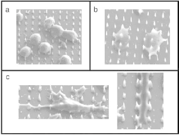
Figure 8.8
Scanning electron micrographs of CHO cells after (a) centrifugation of
cells onto vertically aligned carbon nanofiber array (b) press of vertically aligned
carbon nanofibers into the cells, and (c) culture for 48 h after steps a and b.
either adsorbed or covalently attached plasmid DNA, fiber arrays can be pressed
into (i.e., interfaced with) viable cells (Figure 8.8) on a massively parallel basis
to deliver genetic material or other macromolecules into the cells.
Once interfaced to nanofibered substrates, cells were observed to recover
and proliferate on the fibered substrate and to express the delivered gene for
extended time periods (Figure 8.9). In addition to demonstrating a promising
method for
massively parallel
microinjection of cellular/tissue matrices, the re-
sults of these experiments indicated that plasmid DNA can be expressed within
CHO cells even while covalently tethered to a penetrant nanofiber scaffold.
As these plasmid DNAs are immobilized on the nanofiber, their potential for
genomic insertion is likely reduced, and data indicated that segregation of these
bound plasmids to progeny is rare. Thus, the cells interfaced with the nanofibers
can be provided with expressable, exogenous DNA that is non-inheritable (Fig-
ure 8.10). Furthermore, these plasmids can be physically removed from the cell
along with the nanofiber scaffold, providing a unique strategy for temporal
control of gene expression, the basis of all cellular information processing.
In addition to gene delivery and physical control of gene expression, nano-
fibers may be implemented as electrochemical probes that can be used for
cell-to-chip and chip-to-cell communication, either by direct electrochemi-
cal methods (oxidation/reduction processes) or by electrochemical/electrical/
thermal manipulation of nanofiber-bound molecules, including DNA and