Biomedical Engineering Reference
In-Depth Information
d
n
4
t
3
n
g
|
0
n
3
.
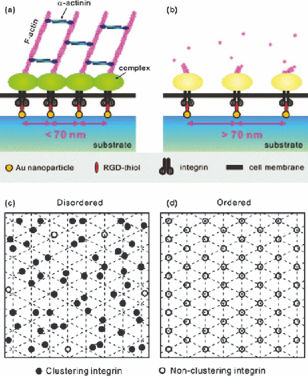
Figure 2.14
Sketch of integrin clustering and subsequent Focal adhesion (FA)
formation regulated by different c(-RGDfK-)-thiol ligand nanopatterns.
The spatial arrangement of the original Au nanopatterns well reflects the
c(-RGDfK-)-thiol ligand lattice and, thus, the final lateral positioning of
single integrins during cell adhesion. (a) A spacing of
o
70 nm between
two neighboring c(-RGDfK-)-thiol ligands results in effective integrin
clustering and FA complex formation, followed by the formation of the
F-actin cytoskeletal network (only some of the intracellular molecules
were depicted here). (b) In contrast, a spacing of 470 nm, as such, results
in neither integrin clustering nor FA complex formation. (c, d) It was
presumed that all integrins that potentially bind c(-RGDfK-)-thiol
ligands over each nanopattern could be classified as clustering integrins
(black disks); non-clustering integrins (white disks) resulted from inter-
distances above the critical value. Even at a global average interligand
spacing of 470 nm, a disordered nanopattern still displayed some clus-
tering integrins, which was not the case for ordered patterns with inter-
ligand spacings of 470 nm.
(Reprinted by kind permission from the American Chemical Society).