Biomedical Engineering Reference
In-Depth Information
Lieber group at Harvard University has reported silicon nanowire field-effect
transistors (NW-FETs) arrays. They showed that simultaneous recordings
from the axon and dendrites of a single neuron were possible with NW-FET
arrays.
13
In addition, neural signals ranging from 0.3mV to 3mV were
recorded from neural circuits in brain slices using a NW-FET array.
14
NW-
FET is a promising sensor that can provide sucient sensitivity with
unprecedented spatial selectivity.
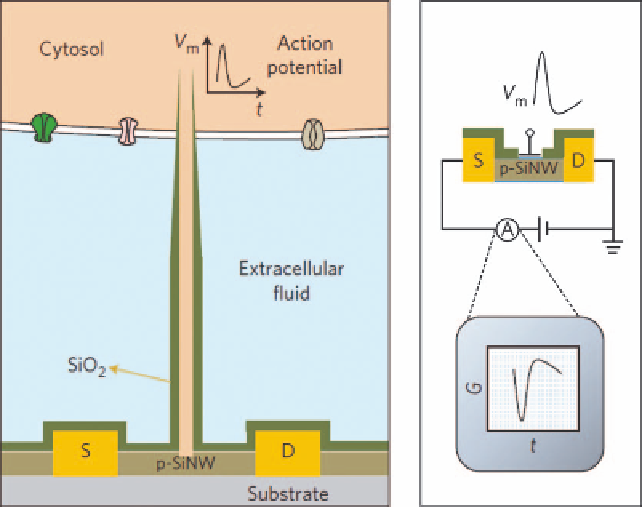
Field-effect transistors (FETs) can also record intracellular electric
potentials. As their performance does not depend on electrode impedance, they
can be made much smaller than micropipettes and microelectrodes. FET arrays
are better suited for multiplexed measurements. SiO
2
nanotubes synthetically
integrated on top of a nanoscale FET penetrate the cell membrane, bringing the
cell cytosol (Figure 4.3) into contact with the FET, which is then able to record
the intracellular transmembrane potential.
15
Branched intracellular nanotube FETs (BIT-FETs) possess a bandwidth
high enough to record fast action potentials even when the nanotube diameter
is reduced to 3 nm, a length scale well below that accessible with other methods.
Studies show that a stable and tight seal forms between the nanotube and cell
d
n
4
t
3
n
g
|
0
n
3
.
Figure 4.3
Schematic diagrams showing (left) a cell coupled to a BIT-FET and the
variation in device conductance G (right) with time, t, during an action
potential V
m
. S and D indicate source and drain electrodes, respectively.
The SiO
2
nanotube connects the cytosol (orange) to the p-type silicon
nanowire FET and, together with the SiO
2
passivation (green), excludes
the extracellular medium (light blue) from the active device channel.
15
(Reprinted by kind permission from Nature Publishing.)