Biology Reference
In-Depth Information
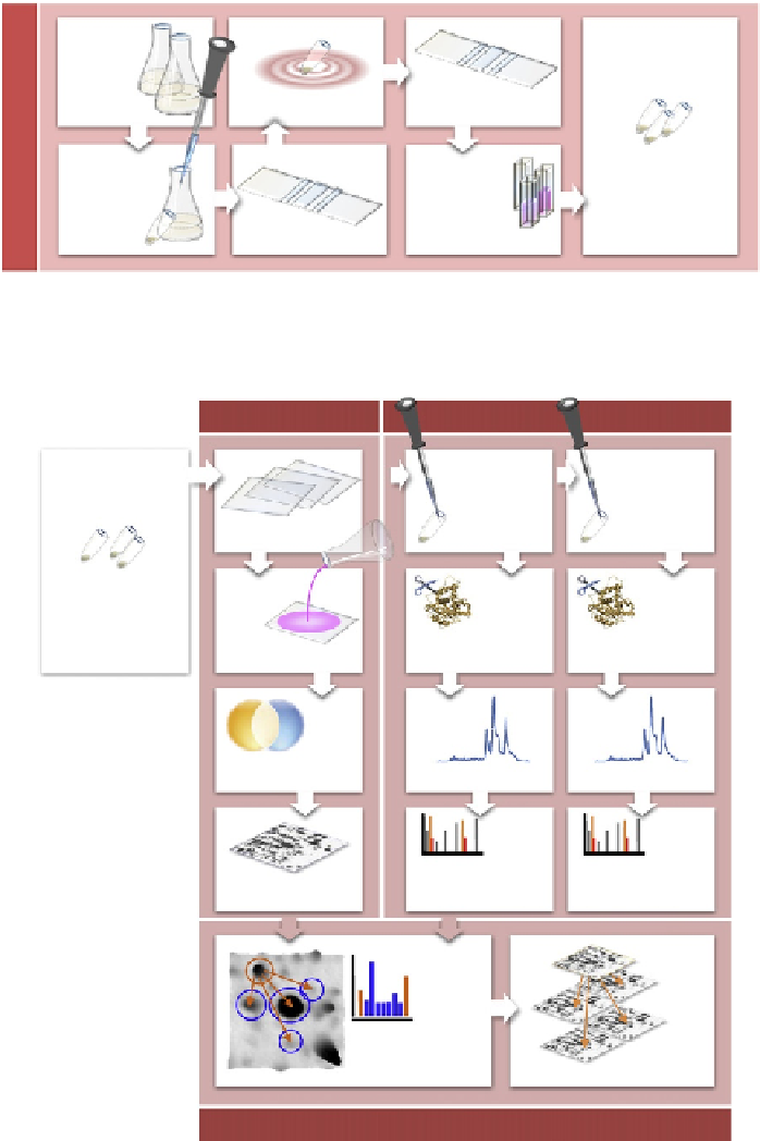
A
Evaluation of cell
disruption efficiency
needed for every specific
experiment!
Dependency of cell
disruption method on
organism and growth state
Integration of data for cell
count calibrated proteins
sample to be used for 2D
gel analysis and for
targeted proteomics
experiment
Standard operation
procedures
Inoculation cell numbers
Cultivation flasks/shaking
system: up-/downscaling
In cell morphology/ growth
state
Cultivation
Cell counting II
Cell desintegration
Cell count
calibrated
protein samples
Protein
content
measurement
with Ninhydrin
Sampling
Cell counting I
Sampling speed
Conditions appropriate for
respective-omics
technique?
Removal of buffers
Suitability of growth media
for subsequent analyses?
Counting chamber
Photographing series of
areas helps to shorten
analysis time
Evaluation of method
advisable
Avoidance of any protein
specific bias
B
2D gels
Calibration of anchor proteins
Spike-in of
heavy
(AQUA)
peptides
Spike-in of
heavy
(QconCAT)
protein
2D gel
electrophoresis
Cell count-
calibrated
protein samples
Tryptic
digestion
QconCAT protein
will be cleaved to
heavy ref peptides
Tryptic
digestion
heavy peptides
remain unchanged
Sequence
unspecific staining
Image
analysis
LCMS precursor ions
LCMS precursor ions
LCMS &
MS/MS
LCMS &
MS/MS
determines
light
peptide/
heavy
peptide
ratios
determines
light
peptide/
QconCAT
peptide
ratios
Image
acquisition
Reference
-based
quantitation of all
proteins
Intergel
calibration
2D gel calibration