Biology Reference
In-Depth Information
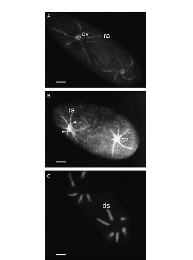
Figure 9.1 Examples of several CVC components of P. tetraurelia visualized by fluores-
cence microscopy. (A) Syb2-GFP stains the CV and radial arms with adjacent smooth
spongiome (confirmed by high-resolution immuno-EM). (B) NSF localized by anti-NSF
antibodies under precautions allowing for the retention of the antigen at the sites of
NSF activity. Note staining of CV and radial arms with adjacent smooth spongiome
(evident from comparison with (A)) and hot spots at the CV/radial arms junctions
(arrowheads). (C) H
þ
-ATPase visualized by the F-subunit (of the catalytic V1 part) as a
GFP-fusion protein reveals its localization to the decorated spongiome (confirmed by
immuno-EM). cv, contractile vacuole; ds, decorated spongiome; ra, radial arm (with
decorated spongiome attached). Bars
¼
10 mm. (A) From
Schilde et al. (2006)
, (B) from
Kissmehl et al. (2002)
, and (C) from
Wassmer et al. (2005)
.
Search WWH ::

Custom Search