Biology Reference
In-Depth Information
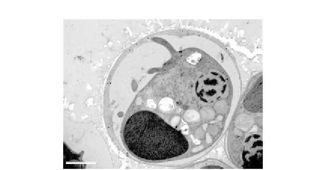
Figure 8.9 Ultrastructure of immature zoospore of C. velia. While both flagella of this
zoospore have already been formed, it remains encysted. Note the large plastid and
numerous cytosolic granules (scale bar
m). Reprinted from
Oborník et al. (2011)
,
Copyright (2011), with permission from Elsevier 2011.
¼
1
m
a unique finger-like projection located close to the root of the shorter fla-
gellum (
Fig. 8.8
), a feature absent from the other ultrastructurally studied
chromerid
V. brassicaformis
(
Oborn´k et al., 2012
), or the closely related
colpodellids (
Leander et al., 2003
). It appears that axonemes are formed
inside the round aflagellar cell, with the flagella being at once ejected to
its surface (
Fig. 8.8
)(
Oborn´k et al., 2011
). It is worth noting that such
an unusual way of flagellar formation has been earlier described in
Plasmo-
dium
(
Briggs et al., 2004; Killick-Kendrick and Peters, 1978
). Moreover, a
structure
resembling the pseudoconoid of
some
colpodellids
and
apicomplexans is also present in
C. velia
(
Oborn´k et al., 2011
).
2.2.
: An alga from the Great Barrier Reef
The second chromerid species known so far was isolated byRobertMoore from
the stony coral
Leptastrea purpurea
in the vicinity of One Tree Island, the Great
Barrier Reef, and was formally described as
V. brassicaformis
(
Oborn´ketal.,
2012
). Both chromerid species have very different morphology and life cycle,
as well as the structure of their plastid genomes and evolutionary tempo of
respective plastid-encoded genes (
Janouˇkovec et al., 2010; Oborn´ketal.,
2011, 2012
).Recent results of a large-scale investigationof chromerid sequences
demonstrated an unexpectedly high abundance of
Vitrella
-like sequences in the
ocean, relatively rare occurrence of those affiliatedwith the genus
Chromera
,and
the presence of several novel chromerid lineages, for which morphological data
have yet to be obtained (
Janouˇkovec et al., 2012a,b
).
V. brassicaformis
Search WWH ::

Custom Search