Biology Reference
In-Depth Information
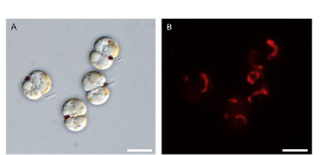
Figure 8.2 Light microscopy of cultured Chromera velia. Coccoid cells after the first
round of cell division (left panel). Autofluorescence of the plastids (right panel) (scale
bar
¼
5
m).
m
discussion (see
Chapter 7
for details). The culture, derived from the origi-
nally isolated strain, is being maintained in our lab for about 8 years as a free-
living full phototroph in a simple algal F2 medium without any external
source of organic carbon. Nonmotile brownish coccoid cells from 5 to
7
m
m in diameter represent the originally isolated stage and also dominate
the culture (
Fig. 8.2
). These vegetative coccoids form colonies where indi-
vidual cells are kept together by some kind of a sticking gel produced by the
alga. In the stationary culture, individual cells as well as colonies of
C. velia
sediment, forming a brownish layer at the bottom of the cultivation flask,
with a tendency to adhere to its surface, suggesting possible endophytic char-
acter of chromerid algae. In addition to the predominant nonmotile coc-
coids, flagellated cells termed zoospores possessing two heterodynamic
flagella were observed in the culture (
Mooreetal.,2008
). As found later
on, formation of zoospores is driven by exposure to light (
Oborn
´
k et al.,
2011; Weatherby et al., 2011
). Since the rhythmicity of zoospore formation
is preserved also under stable light condition, it is likely subject to circadian
clock proteins that have indeed been found in the
C. velia
genome
(Kruˇinsk´ et al., unpublished results). Under culture conditions, the abun-
dance of flagellates is also influenced by salt in the cultivation medium, as
higher salinity of the medium resulted in a lower level of zoospore production
(
Guo et al., 2010
). The function of zoospores in the life cycle of
C. velia
remains to be established. However, when the culture is exposed to spotlight
of high intensity, coccoids massively transform into zoospores, which are
Search WWH ::

Custom Search