Biology Reference
In-Depth Information
2.2.1 Cell polarity of the oocyte and egg
Epithelial cells are polarized along their apicobasal axes with respect to exte-
rior or lumenal environments. These cells may also exhibit polarity with
respect to their orientation within a tissue (i.e., planar cell polarity; reviewed
in
St Johnston and Ahringer, 2010
). Despite their epithelial origin, oocytes
do not have asymmetric epithelial-like cortical polarity. Instead, the oocyte
cortex appears to have mixed apical and basolateral character (
Fig. 4.2
). Pro-
teins normally restricted to the basolateral domains of epithelial cells, Cdh3
(Cadherin 3, type 1, P-cadherin; XB/U-cadherin) and Itgb1 (Integrin, beta
1), are expressed uniformly on the oocyte plasma membrane (
Angres et al.,
1991; M¨ ller et al., 1992, 1993
), as is an apical protein, Atypical protein
kinase C (Prkci/aPKC/protein kinase C, iota;
Nakaya et al., 2000
). The bas-
olateral proteins are internalized during oocyte maturation and reexpressed
in newly formed cleavage furrows after fertilization. Interestingly, these new
membranes form the basolateral domains of blastomeres, whereas inherited
egg membrane and cortex become apical membrane (
M
¨
ller and Hausen,
1995
), to be inherited by superficial epithelial cells of the blastula.
Membrane trafficking in oocytes occurs uniformly but becomes polar-
ized to new membrane upon first cleavage (
Roberts et al., 1992
). Addition-
ally, proteins synthesized during oogenesis and stored in post-Golgi vesicles
are only competent for insertion into newly formed, basolateral membrane
during cleavage, although the mechanism for this specificity is not known
(
Roberts et al., 1992
). Despite these early functional differences between
egg and newmembrane domains in early blastomeres, fully mature epithelial
polarity is not evident until the morula stages (
M
¨
ller and Hausen, 1995
).
Oocyte
Egg
Embryo
Prkci
Chd1
,
ltgb1
,
Llgl2-GFP
Chd1
,
ltgb1
,
Prkci
,
Llgl2-GFP
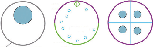
Figure 4.2 Distribution of cell polarity proteins during the oocyte-to-embryo transition
in Xenopus. In the oocyte, apical and basolateral cell polarity proteins are uniformly dis-
tributed in the membrane. Following oocyte maturation, apical proteins (e.g., Prkci) are
enriched animally, many basolateral proteins are internalized (e.g., Cdh1, Itgb1). After
fertilization and cleavage of the egg, the egg membrane acquires apical character
and newly made internal membrane reexpress basolateral markers.

Search WWH ::

Custom Search