Biology Reference
In-Depth Information
A
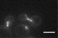
S. cerevisiae
B
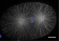
C. elegans
C
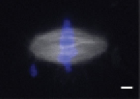
X. laevis
D
H. sapiens
E
A. thaliana
Figure 3.6 Spindle morphology has changed concomitantly with the diversification of
eukaryotic species and with specialization of cells within an organism. All scale bars
5 mm. In the schematics, spindle MTs are indicated in gray, kinetochore fiber MTs in
red, and astral MTs in green. (A) Spindles in budding yeast contain one MT per kinet-
ochore fiber in a closed mitosis, with few astral MTs that extend to the cell cortex.
(B) The C. elegans one-cell embryo spindle contains large astral arrays of MTs that extend
to both chromosomes and the cortex. Single kinetochore fiber MTs bind at intervals
along the length of the entire holocentric chromosome, which contains kinetochore
features throughout. (C) X. laevis meiotic spindles formed in cytoplasmic egg extracts
contain many short MTs that are held together by motors and cross-linking activity
into a large tiled array. (D) Somatic spindles in HeLa cells contain dominant and ro-
bust kinetochore fibers with a moderate number of astral MTs which contact the cortex
Continued


Search WWH ::

Custom Search