Biomedical Engineering Reference
In-Depth Information
TABLE 12.1 General Phenotypic Changes Typically Investigated Using Image-Based High Content Screening Assays
Imaging Assays
Investigating . . .
Descriptive Examples
Literature Examples
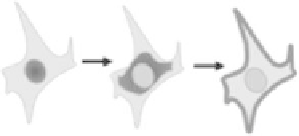
(Sub-)cellular location
Fluorescent marker located in nucleus,
cytoplasm, perinuclear region, or
cell membrane
Estrogen receptor nuclear
translocation [17]
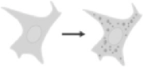
Spatial distribution
Homogeneous vs. punctuate
distribution; long vs. short fibers;
large vs. small vesicles
-arrestin-based GPCR
redistribution assay
(TransFluor) [19]; POD
nuclear foci assay [20]
Co-localization
Fluor 1-labeled protein co-localized
with fluor 2-labeled cell organelle;
two proteins labeled with different
fluorophores co-localized with each
other
Lipid storage vesicles [21]
(
continued
)