Biomedical Engineering Reference
In-Depth Information
temperature. Also, Ca/P ratio of less than 1.0 is not biomedically important. It
needs to be pointed out here that a number of literature reports has emphasized
that nonstoichiometric HAp promotes better osteoconduction property
12
.
Various attempts have been made to experimentally assess the cytocompati-
bility of CaP-based materials. For example, Suzuki et al.
13
showed that serum pro-
tein adsorbed on the surface of TCP-HAp ceramics appears to be effective in
preventing cell rupture by functioning as an intermediate layer that prevents
direct contact between cells and the unstable surface of the materials.
Chen et al.
14
investigated the bone bonding mechanism of crystalline HAp
in vivo
. It is reported that initially, a layer of amorphous HAp was formed on the
HAp implant surface and a bone like apatite layer formed after three months,
in between the implant and bone tissue. At a later stage (after six months), direct
bone-HAp implant contact is established and collagen fi ber enters inside the im-
plant material. Therefore, the interface region shows good mechanical strength
with the new bone apposition.
Xin et al.
15
compared the CaP formation behavior of few bioceramics
in vitro
(simulated body fl uid) and
in vivo
. The investigated bioceramics include
sintered porous solids, including bioglass, glass-ceramics, hydroxyapatite,
α
-
tricalcium phosphate and
-tricalcium phosphate. The presence of octacalcium
phosphate was observed on all types of bioceramic surfaces
in vitro
and
in vivo
,
except on
β
-TCP. They concluded that Ca-P formation on bioceramic surfaces is
more diffi cult
in vivo
than
in vitro
.
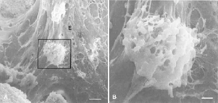
In an interesting study, Dong et al.
16
investigated the effect of low pressure
during culture on the bone formation of osteoblast/porous hydroxyapatite com-
posite
in vivo
. Scanning electron microscopy (SEM) observations (Figure 3.3)
β
Figure 3.3.
A) SEM images of cross-sections of osteoblast-HAp composites after two weeks
of implantation. The pore surface is covered by round cells as well as collagenous extracellular
matrix. E indicates collagenous extracellular matrix. Bar = 37.5 mm. B) Higher magnifi cation of
the large rectangular area in A. Round cells of active osteoblast, can be seen on the surface of
HAp. Bar = 5
m
16
.
μ
Search WWH ::

Custom Search