Biomedical Engineering Reference
In-Depth Information
100
µ
m
-
EF
+
no field
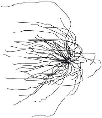
Figure 18.4.
Cathodal turning of axons from embryonic
Xenopus
spinal cord neurons, modi-
fi ed from (McCaig et al. 2005). Used with permission of The American Physiological Society.
from PC12 turn towards the anode (Cork et al. 1994). Therefore, neuronal cell
type, EF strength and extracellular matrix proteins infl uence the behavior of
neurons when grown in an EF. Furthermore, evidence shows that hippocampal
neurons and astrocytes align perpendicularly to electric fi elds
in vitro
(Rajnicek
el al. 1992 ; Alexander et al. 2006 ).
Thus, the infl uence an EF can have on neurites is dependent on EF strength,
time of exposure, nerve cell type, the charge on the substratum (on which the cells
are cultured), and for axonal turning, if the neuronal projection is axonal or den-
dritic (McCaig et al. 2005). These variables create a complicated puzzle when
determining the cellular mechanics that produce responses to EFs.
18.6.2 Cellular Level Changes Due to EFs
It has been proposed that neural growth cone guidance or galvanotaxis in an EF
is due to an accumulation or concentration of receptor and voltage gated chan-
nels in the membrane facing the direction of movement or turning (Figure 18.5A).
The receptors and channels involved are similar to those involved in chemotropic
guidance. For example, in neurons, poly-saccharide-binging plant lectins recep-
tors, such as, concanavalin A receptor (Patel and Poo 1982) and acetylcholine
receptors (AChRs) (Poo 1981; Stollberg and Fraser 1988) are asymmetrically dis-
tributed due to an EF. Similarly, in corneal epithelial cells and fi broblasts, epider-
mal growth factor rector (EGFR) becomes unequally distributed (McCaig et al.
2005). These receptors were shown to accumulate, in an EF, on areas of the cell
membrane facing the cathode.


Search WWH ::

Custom Search